Non Plus image gallery

153 images of radar screens, particle tracks, cells and embryos, collected for the Non Plus collective. Everything here is public domain or Creative Commons; each image lists its author and licence.

Click Download to save an image, or follow Full resolution for the original at its source. If you use an image, credit it as its caption says.

Radar & scopes

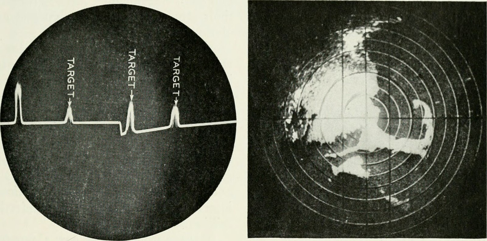
Bell telephone magazine (1945) (14569797239)
1 Bell telephone magazine (1945) (14569797239)
Internet Archive Book Images · Public domain · sourceDownloadFull resolution (2046×1012) ↗
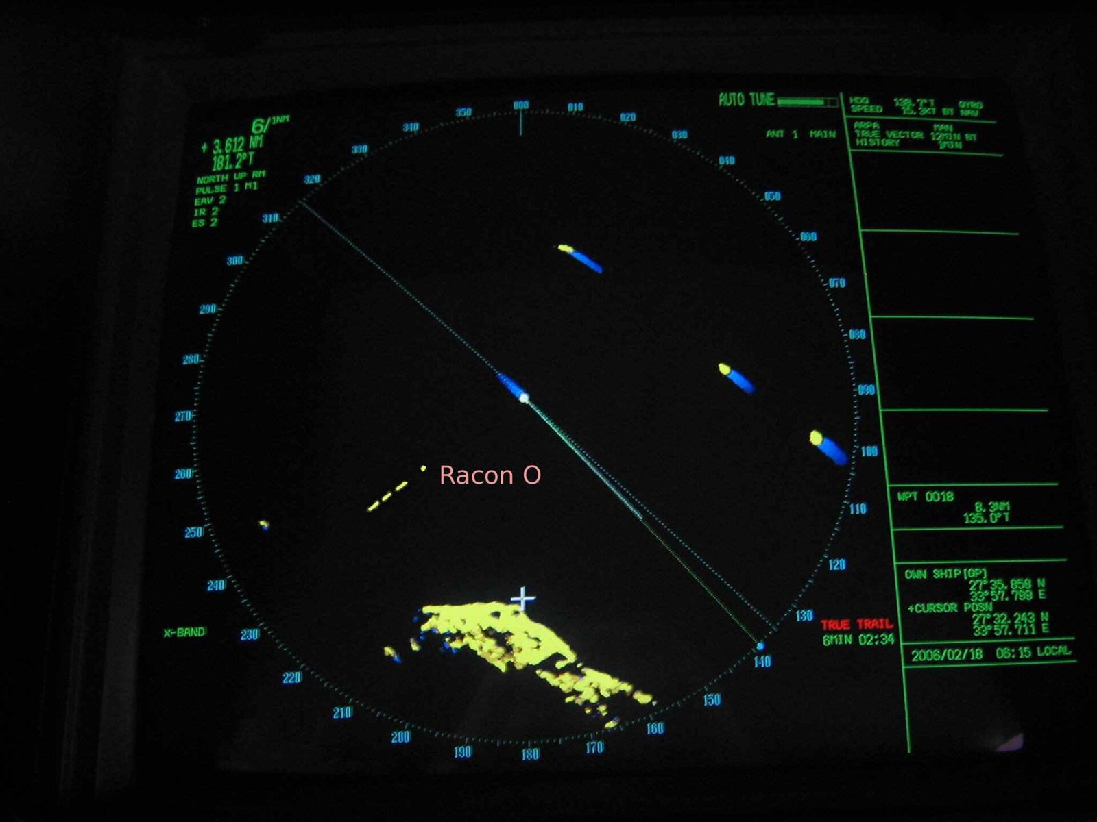
Radar Racon O
2 Radar Racon O
Clipper · CC BY-SA 3.0 · sourceDownloadFull resolution (2048×1536) ↗
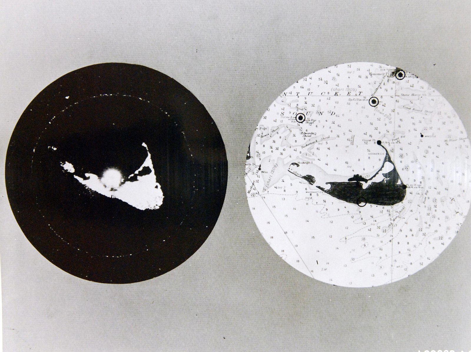
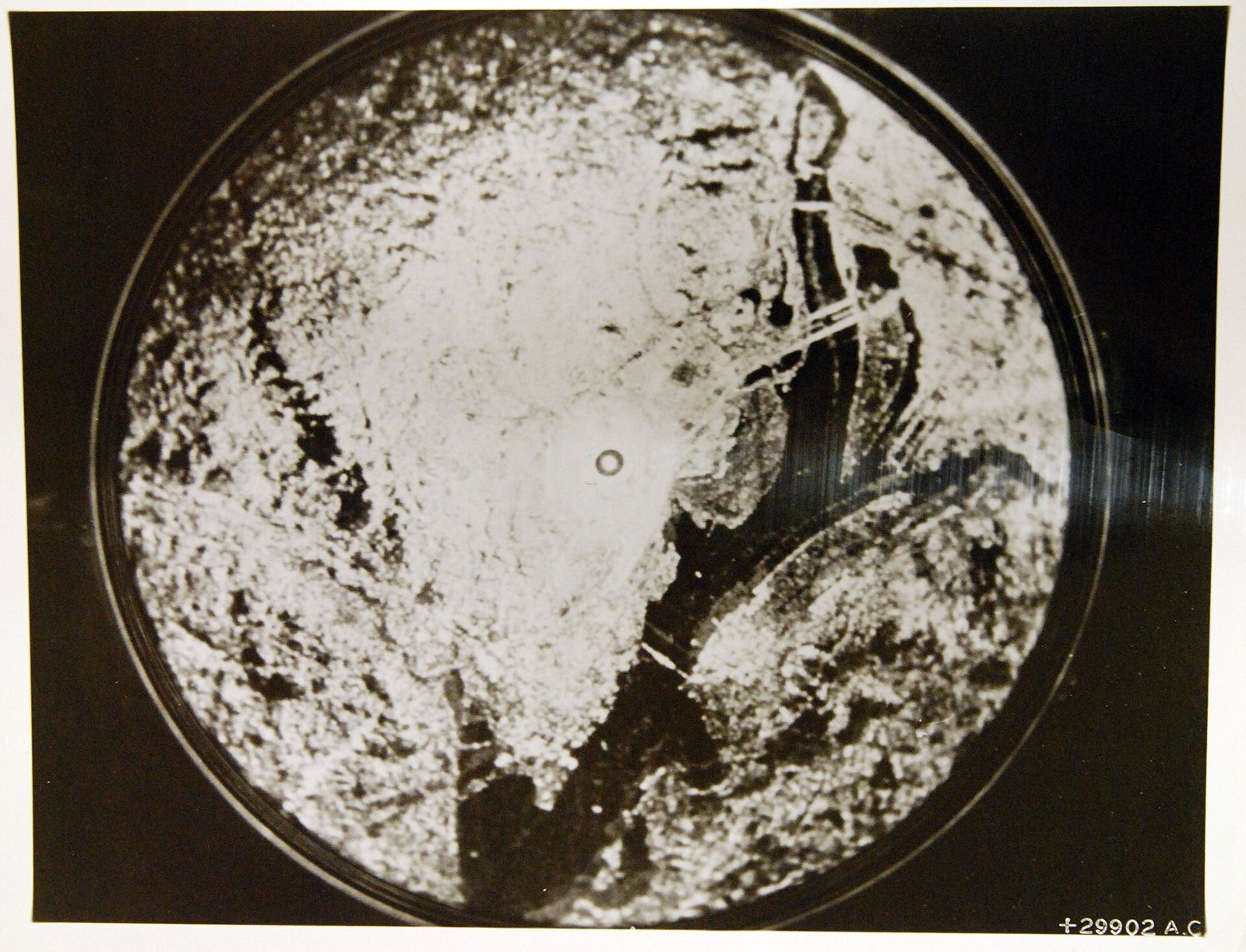
WWII comparison of aerial radar scope photograph of Nantucket with actual chart of island (38628763390)
3 WWII comparison of aerial radar scope photograph of Nantucket with actual chart of island (38628763390)
National Museum of the U.S. Navy · Public domain · sourceDownloadFull resolution (3031×2268) ↗
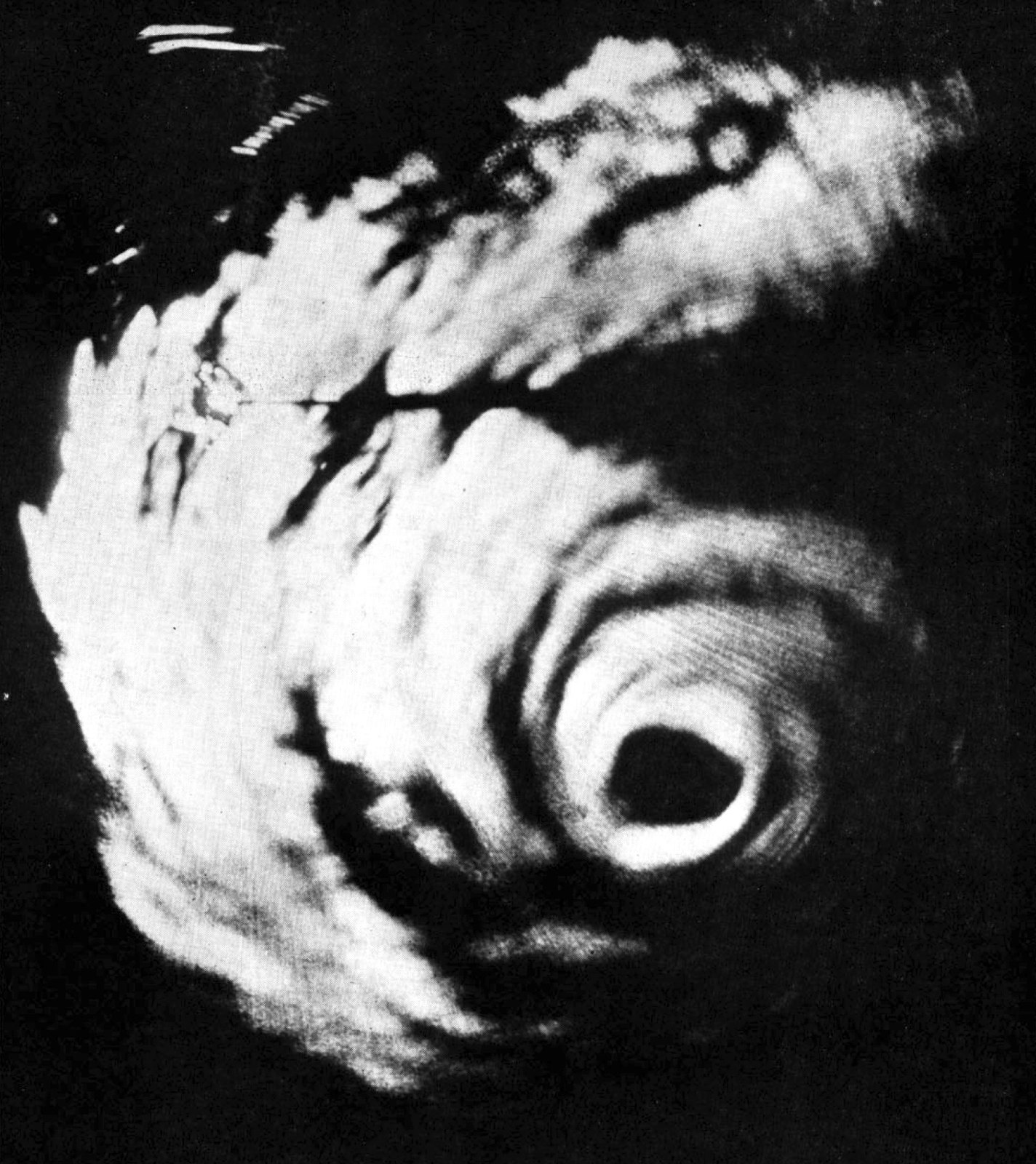
Hurricane Gracie on radar scope in September 1959
4 Hurricane Gracie on radar scope in September 1959
PH2 B.J. Dalma, USN · Public domain · sourceDownloadFull resolution (1515×1702) ↗
Night radar scope image of Potomac approach to Washington, WWII (39543289985)
5 Night radar scope image of Potomac approach to Washington, WWII (39543289985)
National Museum of the U.S. Navy · Public domain · sourceDownloadFull resolution (3131×2392) ↗
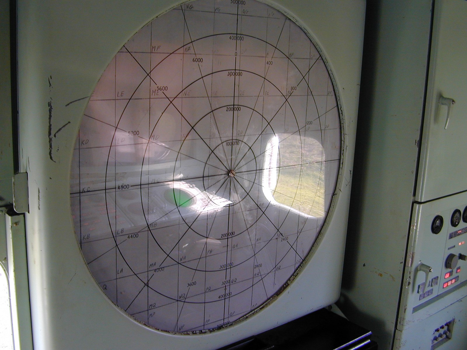
Radar Scope (1961969)
6 Radar Scope (1961969)
Dawn Endico from Menlo Park, California · CC BY-SA 2.0 · sourceDownloadFull resolution (1600×1200) ↗
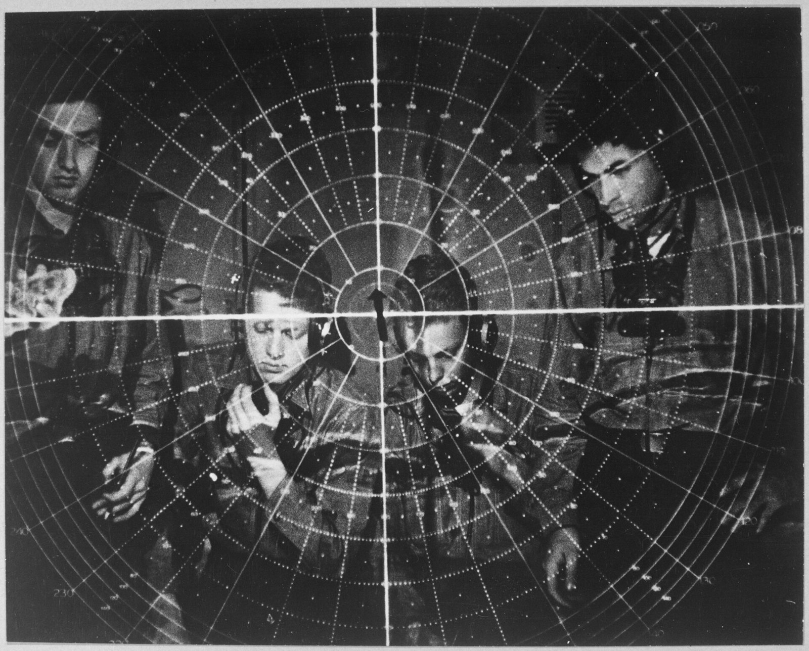
Radar scope. - NARA - 520692
7 Radar scope. - NARA - 520692
Unknown authorUnknown author or not provided · Public domain · sourceDownloadFull resolution (3000×2415) ↗
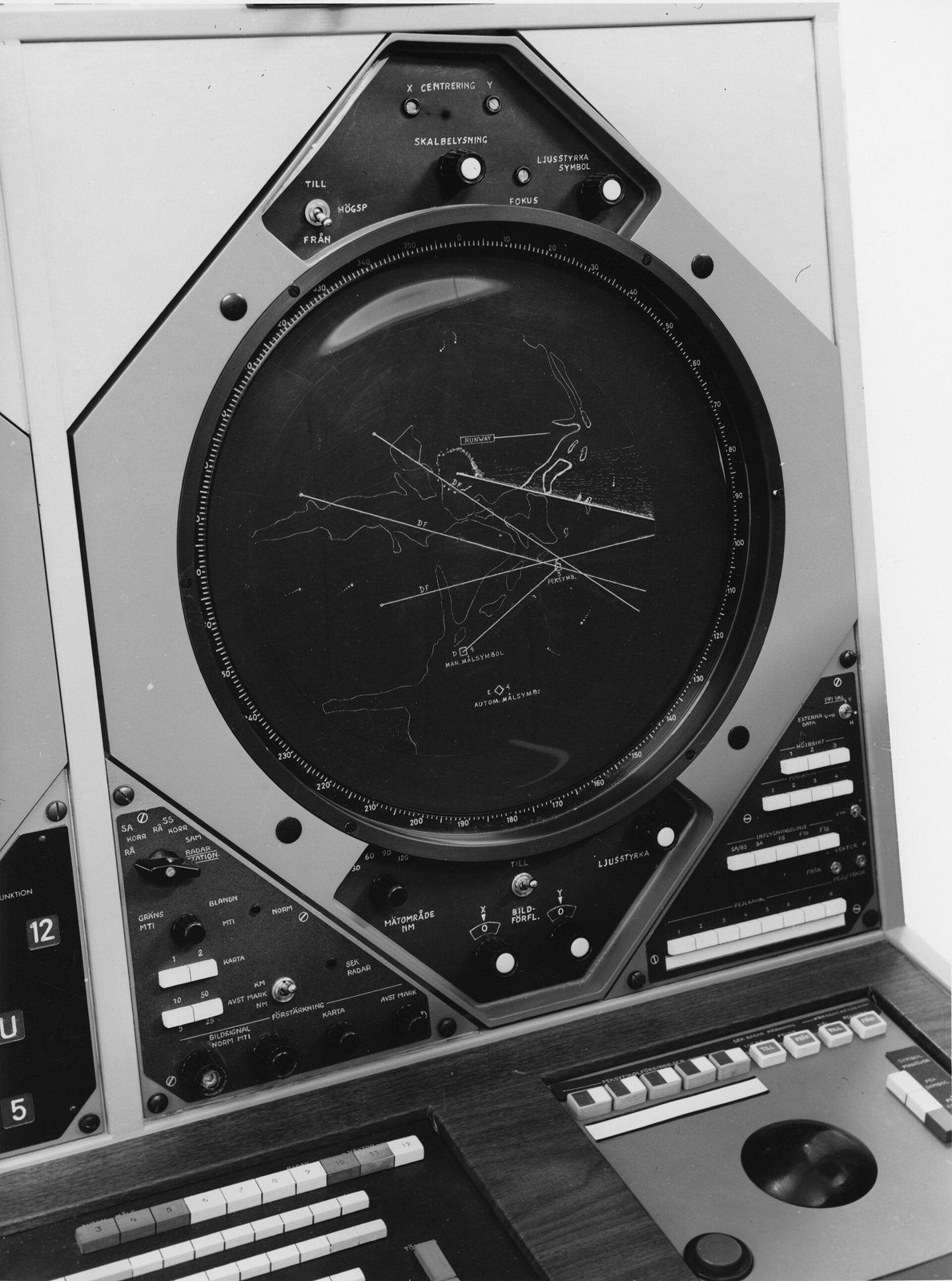
SR&T Plan position indicator
8 SR&T Plan position indicator
Tekniska museet · Public domain · sourceDownloadFull resolution (2634×3543) ↗
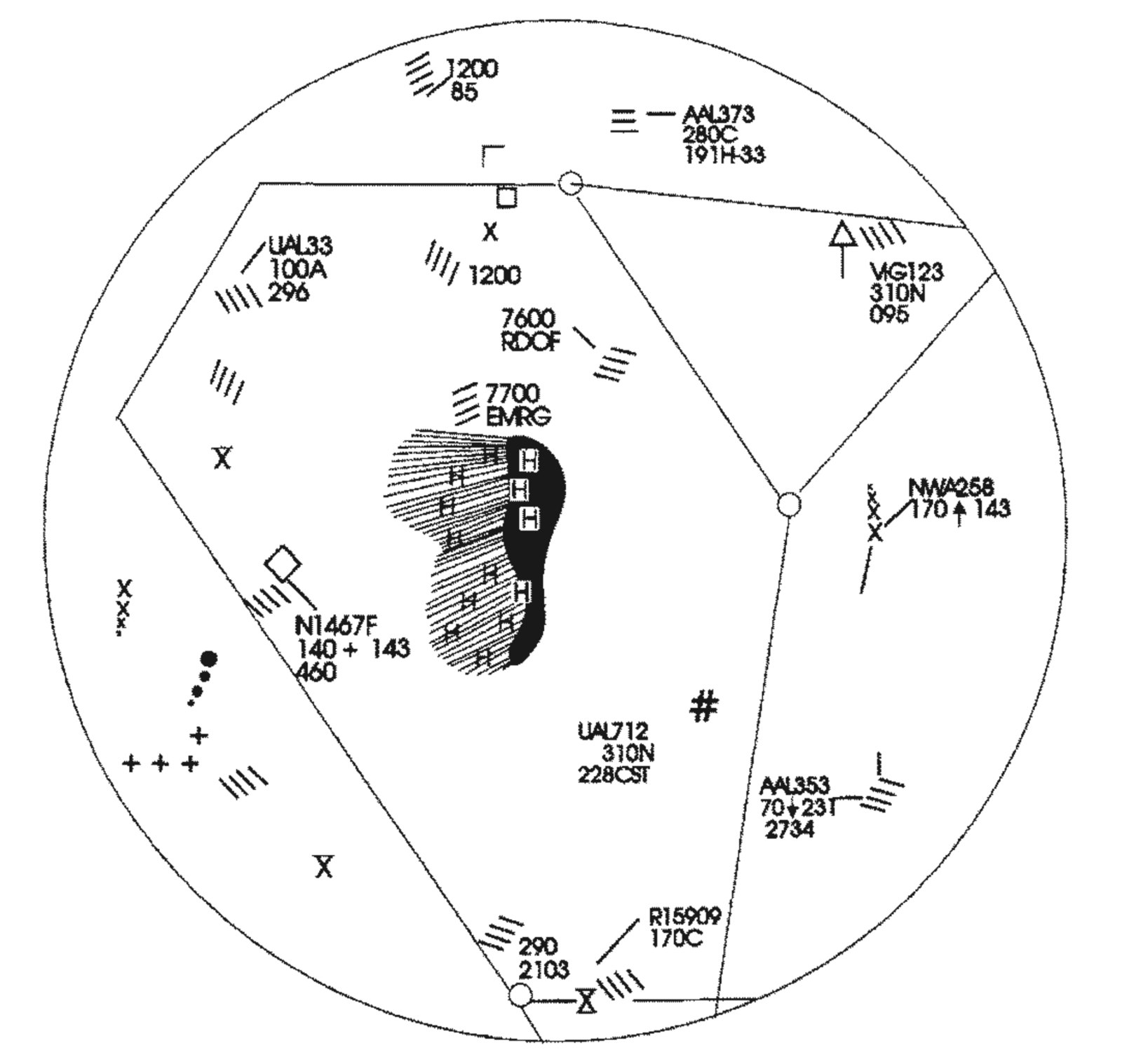
Scope of an ARTCC controller
9 Scope of an ARTCC controller
Federal Aviation Administration · Public domain · sourceDownloadFull resolution (1974×1872) ↗
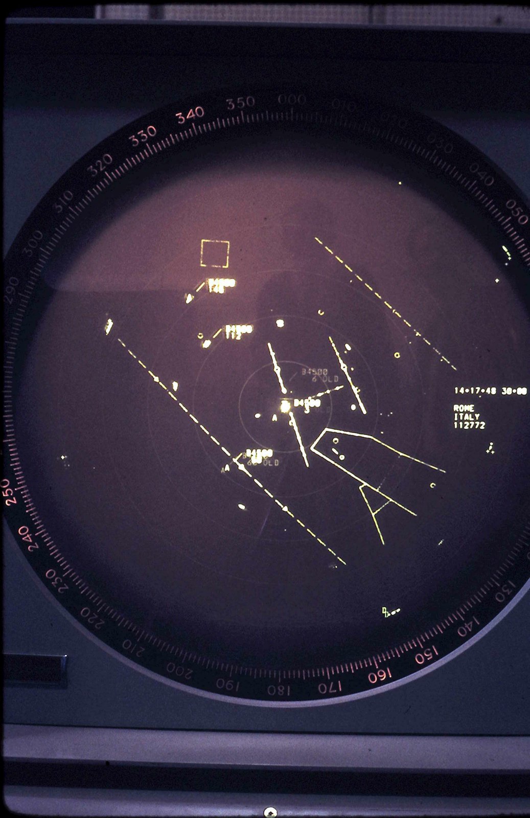
Air Traffic Control Display
10 Air Traffic Control Display
Dms489 · CC BY 3.0 · sourceDownloadFull resolution (1680×2592) ↗
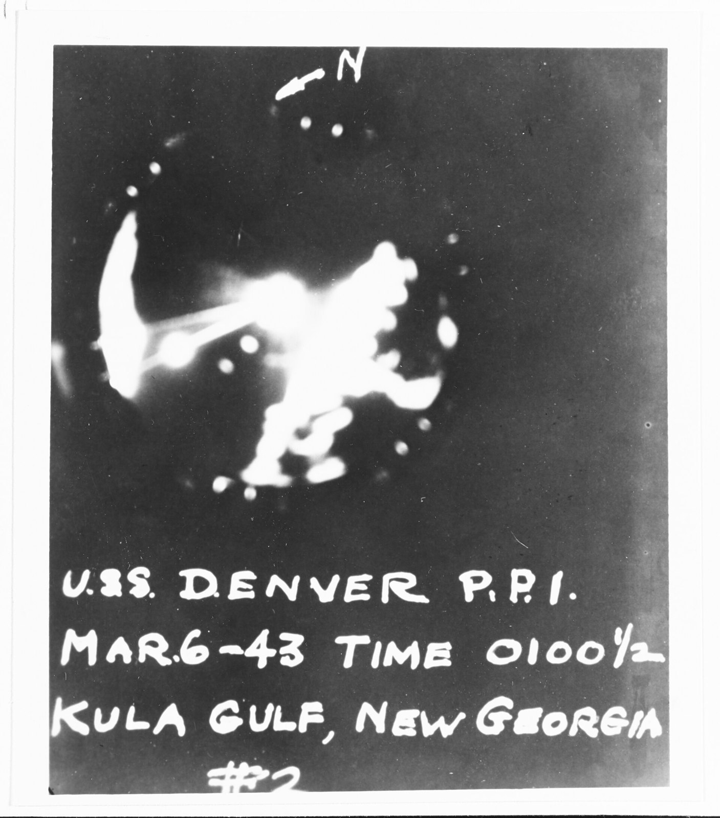
BlackettRadar1
11 BlackettRadar1
Unknown · Public domain · sourceDownloadFull resolution (3156×3588) ↗
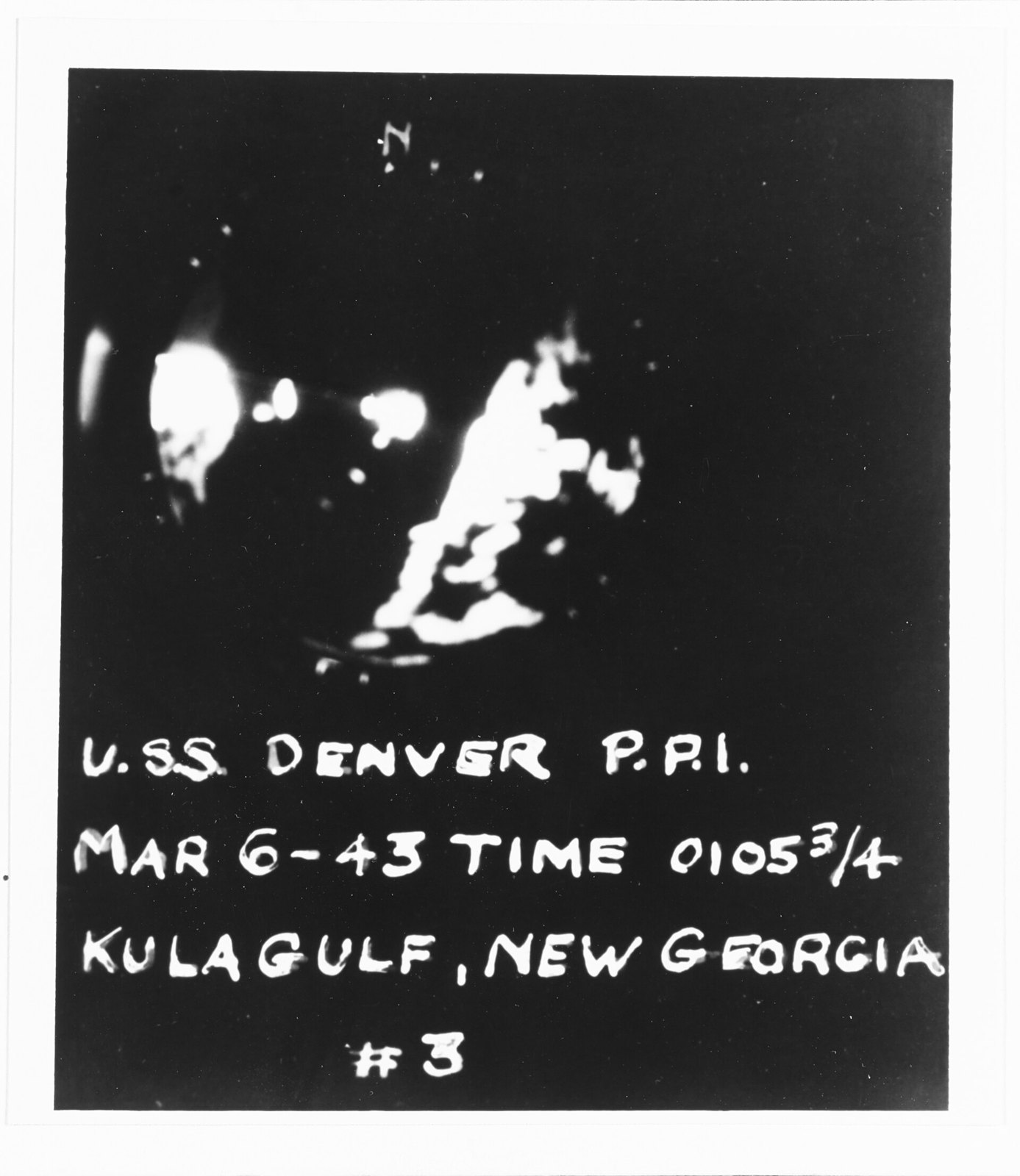
BlackettRadar2
12 BlackettRadar2
Unknown · Public domain · sourceDownloadFull resolution (3108×3586) ↗
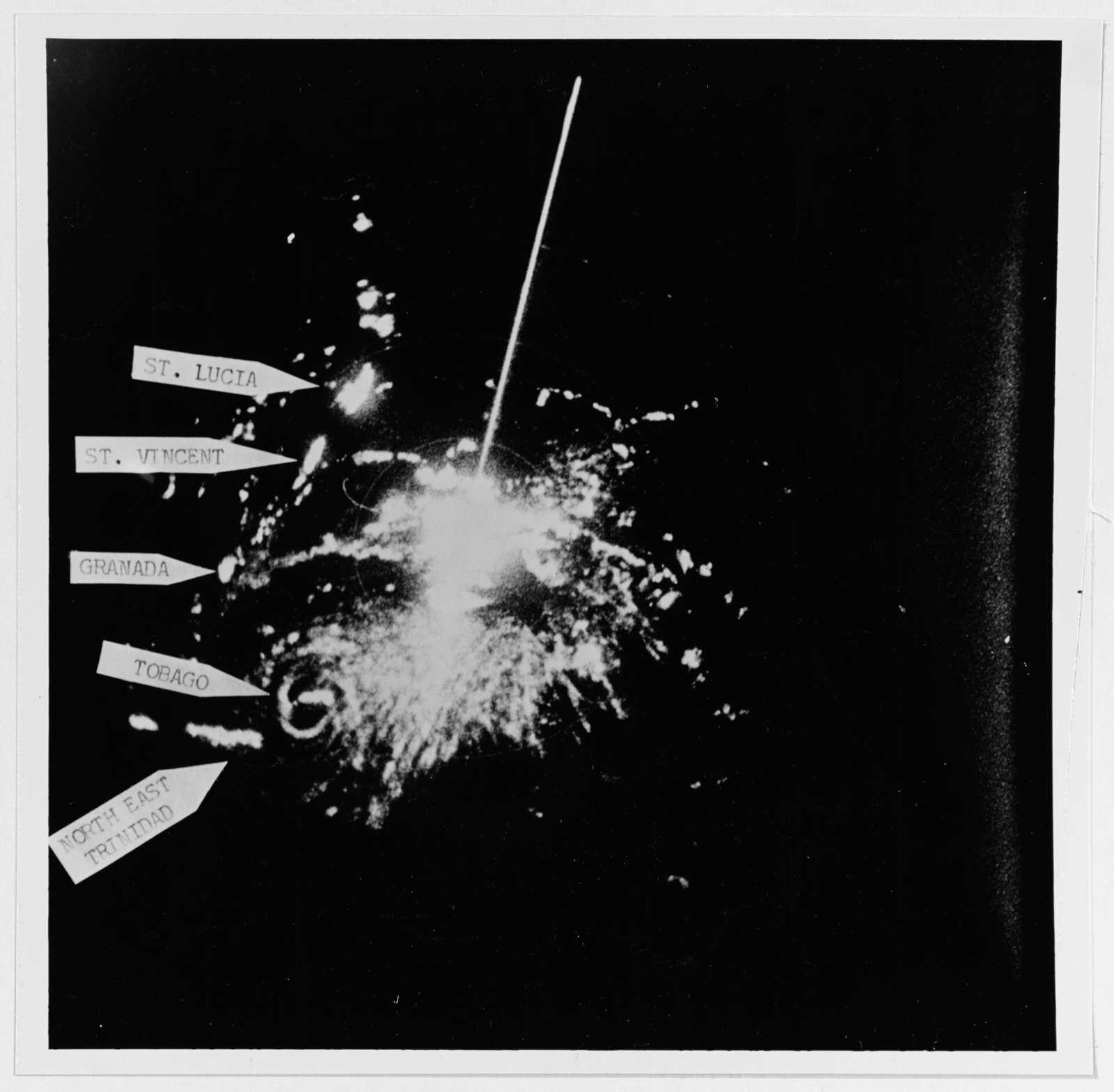
Hurricane Flora over Tobago on radar 1963-09-30
13 Hurricane Flora over Tobago on radar 1963-09-30
U.S. Navy, National Archives · Public domain · sourceDownloadFull resolution (5098×4998) ↗
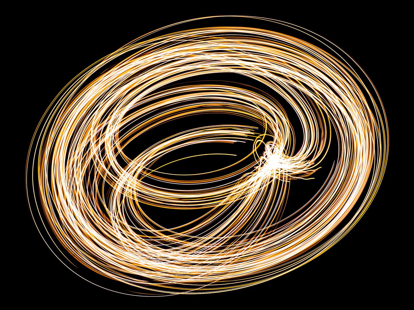
Lissajous-Figur -- 2020 -- 7765
14 Lissajous-Figur -- 2020 -- 7765
Dietmar Rabich · CC BY-SA 4.0 · sourceDownloadFull resolution (5254×3941) ↗
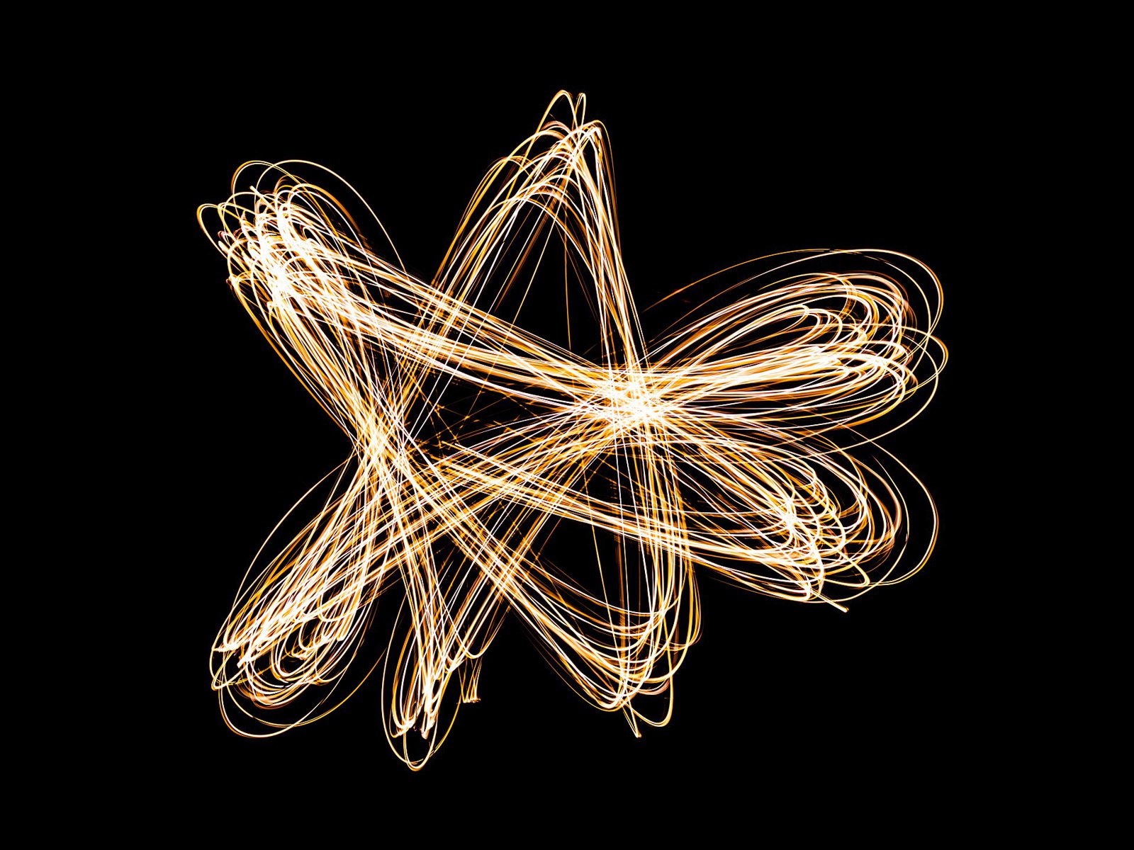
Lissajous-Figur -- 2020 -- 7766
15 Lissajous-Figur -- 2020 -- 7766
Dietmar Rabich · CC BY-SA 4.0 · sourceDownloadFull resolution (5357×4018) ↗
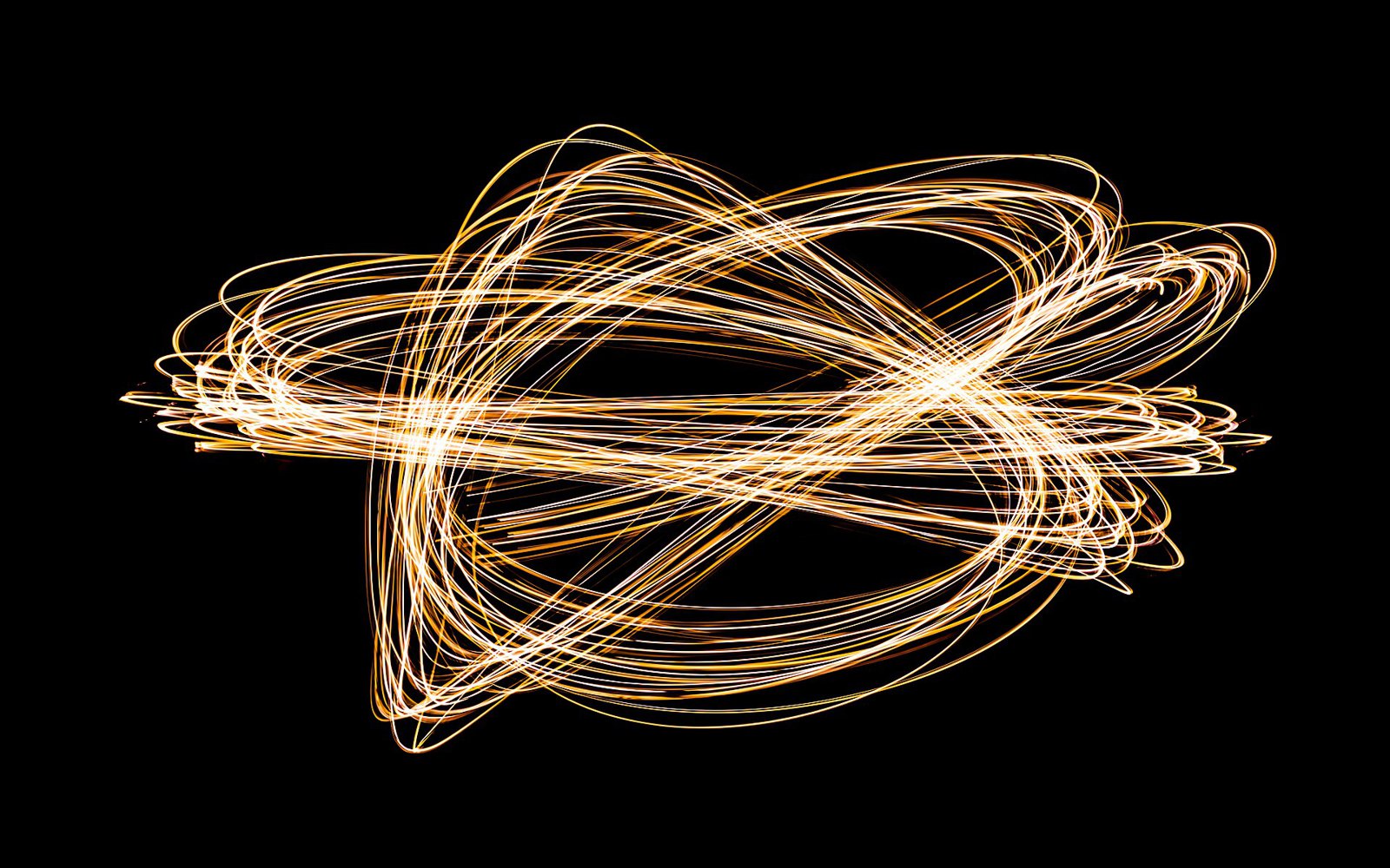
Lissajous-Figur -- 2020 -- 7767
16 Lissajous-Figur -- 2020 -- 7767
Dietmar Rabich · CC BY-SA 4.0 · sourceDownloadFull resolution (6341×3963) ↗
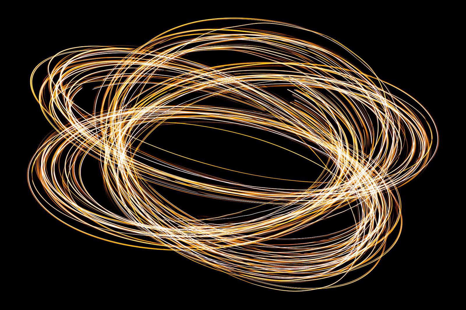
Lissajous-Figur -- 2020 -- 7768
17 Lissajous-Figur -- 2020 -- 7768
Dietmar Rabich · CC BY-SA 4.0 · sourceDownloadFull resolution (6073×4049) ↗
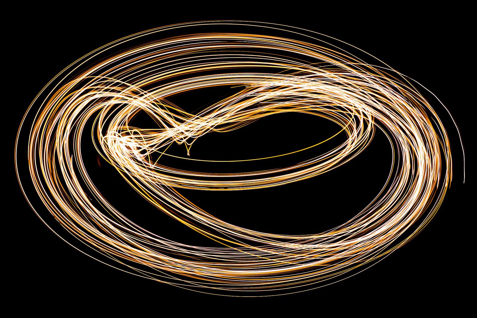
Lissajous-Figur -- 2020 -- 7769
18 Lissajous-Figur -- 2020 -- 7769
Dietmar Rabich · CC BY-SA 4.0 · sourceDownloadFull resolution (6128×4085) ↗
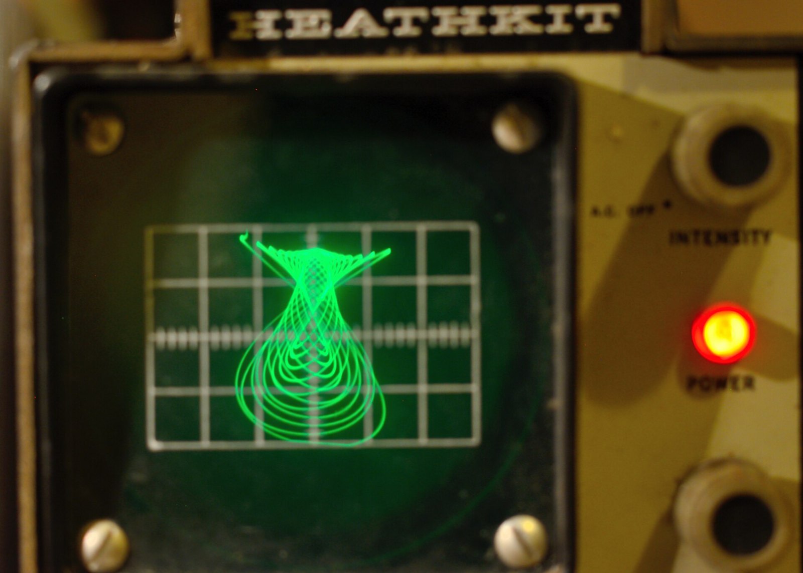
Lissajous figure
19 Lissajous figure
Greg Francke · CC BY-SA 4.0 · sourceDownloadFull resolution (2048×1463) ↗

Particle tracks
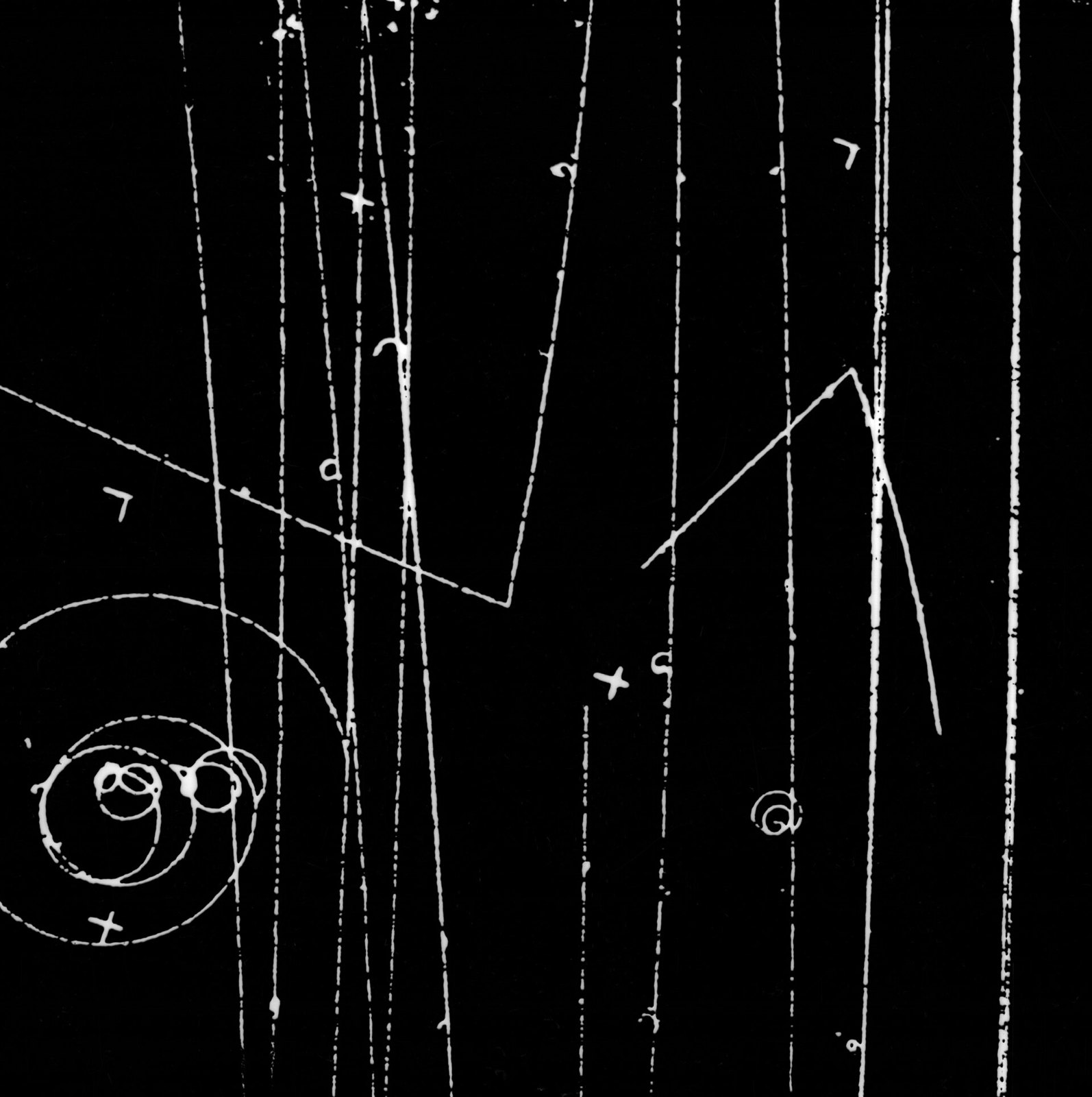
Bubble chamber event, formula demonstration. Photograph taken August 26, 1957. Bubble Chamber-315A - DPLA - aace197da4debc8b3ed9548c646e156c
20 Bubble chamber event, formula demonstration. Photograph taken August 26, 1957. Bubble Chamber-315A - DPLA - aace197da4debc8b3ed9548c646e156c
Department of Energy. Lawrence Berkeley National Laboratory. Public Affairs Department. Strategic Resources Office. Photography Services. 2012 · Public domain · sourceDownloadFull resolution (3200×3212) ↗
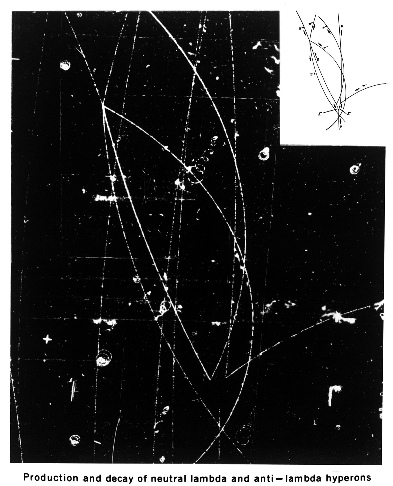
Bubble chamber event. Decay of neutral lambda, anti-lambda hyperons. Photograph circa July 1959. Bubble Chamber-772A - DPLA - 5141859e360581251906c073668c70de
21 Bubble chamber event. Decay of neutral lambda, anti-lambda hyperons. Photograph circa July 1959. Bubble Chamber-772A - DPLA - 5141859e360581251906c073668c70de
Department of Energy. Lawrence Berkeley National Laboratory. Public Affairs Department. Strategic Resources Office. Photography Services. 2012 · Public domain · sourceDownloadFull resolution (2430×3000) ↗
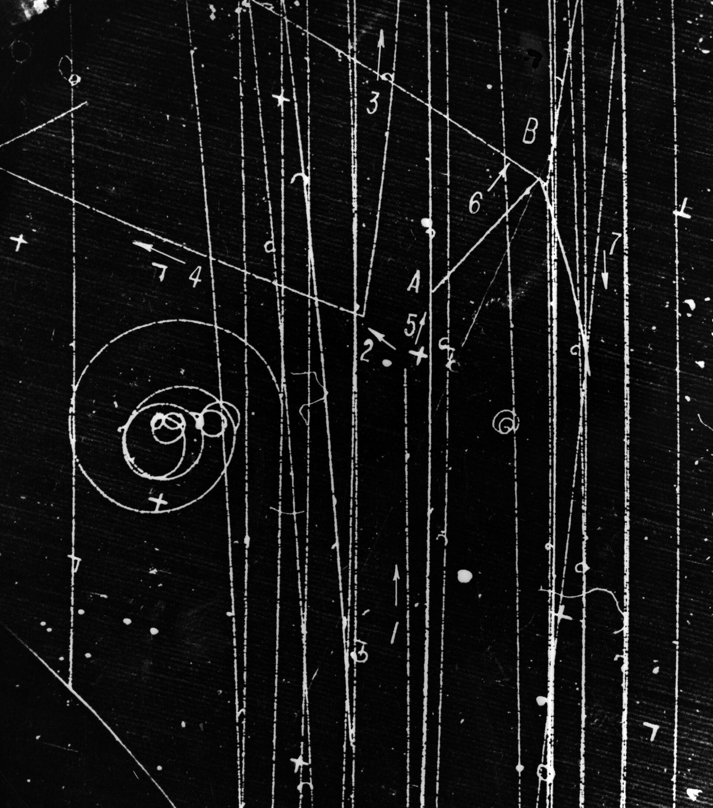
Bubble chamber event. Photograph taken December 16, 1958. Bubble Chamber-315B - DPLA - 57790979981199894c1e2fa1b4bce5e8
22 Bubble chamber event. Photograph taken December 16, 1958. Bubble Chamber-315B - DPLA - 57790979981199894c1e2fa1b4bce5e8
Department of Energy. Lawrence Berkeley National Laboratory. Public Affairs Department. Strategic Resources Office. Photography Services. 2012 · Public domain · sourceDownloadFull resolution (3529×4000) ↗
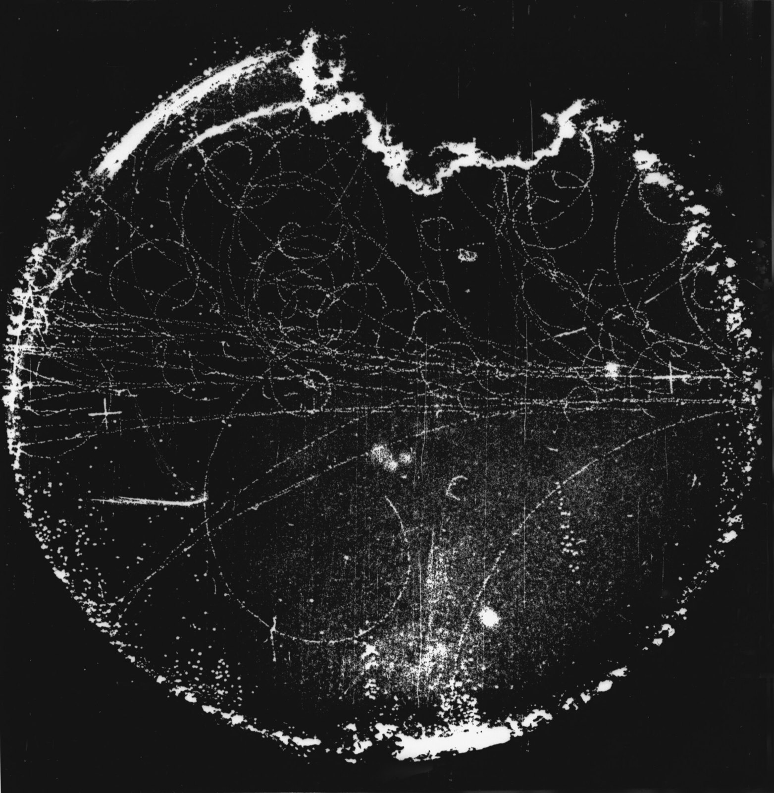
Synchrotron background test in 4-inch bubble chamber. Photograph taken July 27, 1955. Bubble Chamber-27 - DPLA - b6d5a3ec40b03d8ae7481eb9912553ff
23 Synchrotron background test in 4-inch bubble chamber. Photograph taken July 27, 1955. Bubble Chamber-27 - DPLA - b6d5a3ec40b03d8ae7481eb9912553ff
Department of Energy. Lawrence Berkeley National Laboratory. Public Affairs Department. Strategic Resources Office. Photography Services. 2012 · Public domain · sourceDownloadFull resolution (2170×2222) ↗
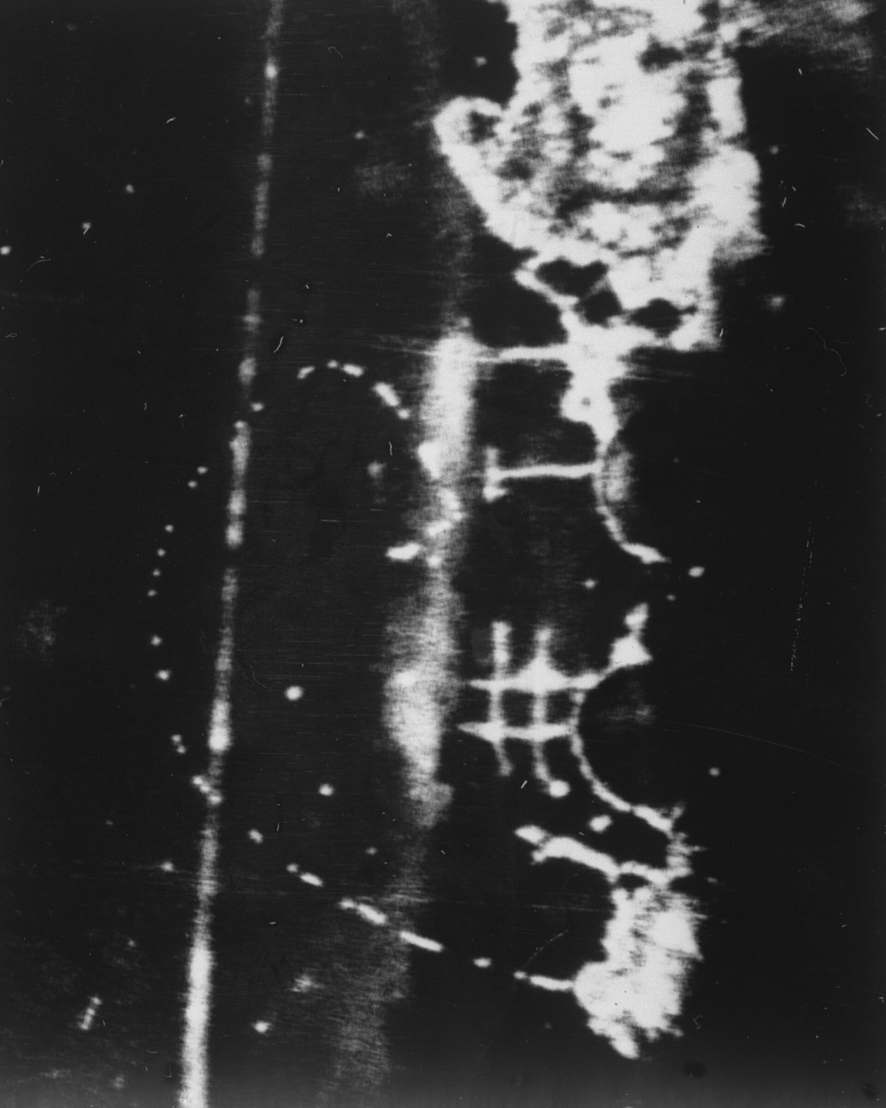
First track in deuterium. Photograph taken October 1955. Bubble Chamber-46 - DPLA - d553c1730076c531c844c8e5e1de1e6c
24 First track in deuterium. Photograph taken October 1955. Bubble Chamber-46 - DPLA - d553c1730076c531c844c8e5e1de1e6c
Department of Energy. Lawrence Berkeley National Laboratory. Public Affairs Department. Strategic Resources Office. Photography Services. 2012 · Public domain · sourceDownloadFull resolution (2400×3000) ↗
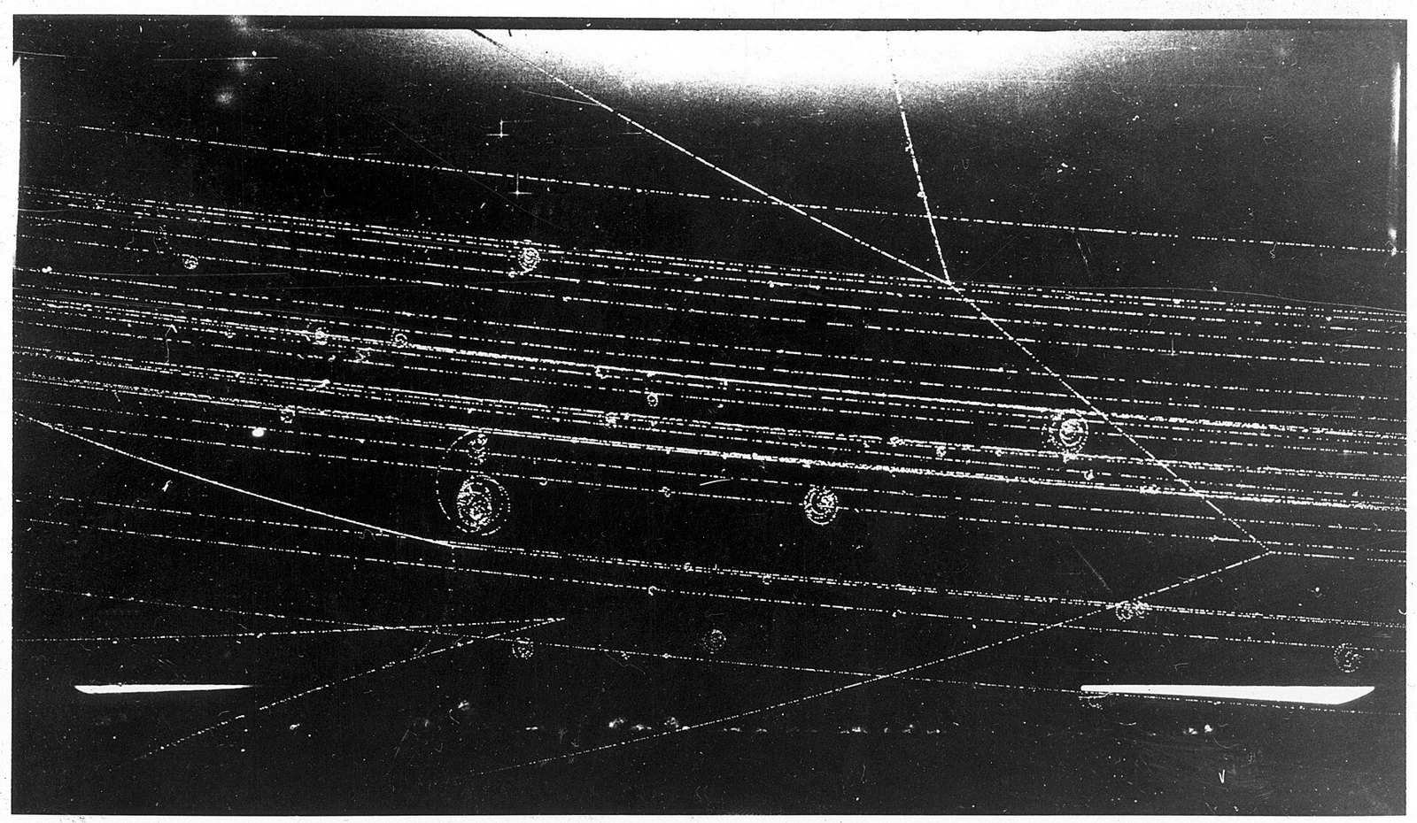
HD.6D.680 (12000818023)
25 HD.6D.680 (12000818023)
ENERGY.GOV · Public domain · sourceDownloadFull resolution (3200×1860) ↗
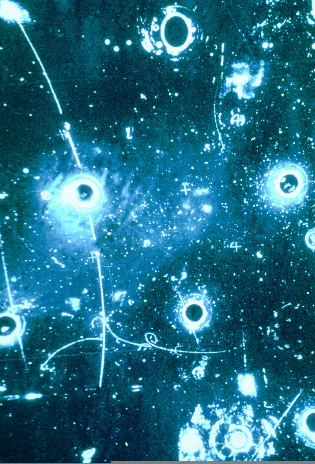
Leptonic event in Gargamelle bubble chamber
26 Leptonic event in Gargamelle bubble chamber
CERN · CC BY 4.0 · sourceDownloadFull resolution (1021×1506) ↗
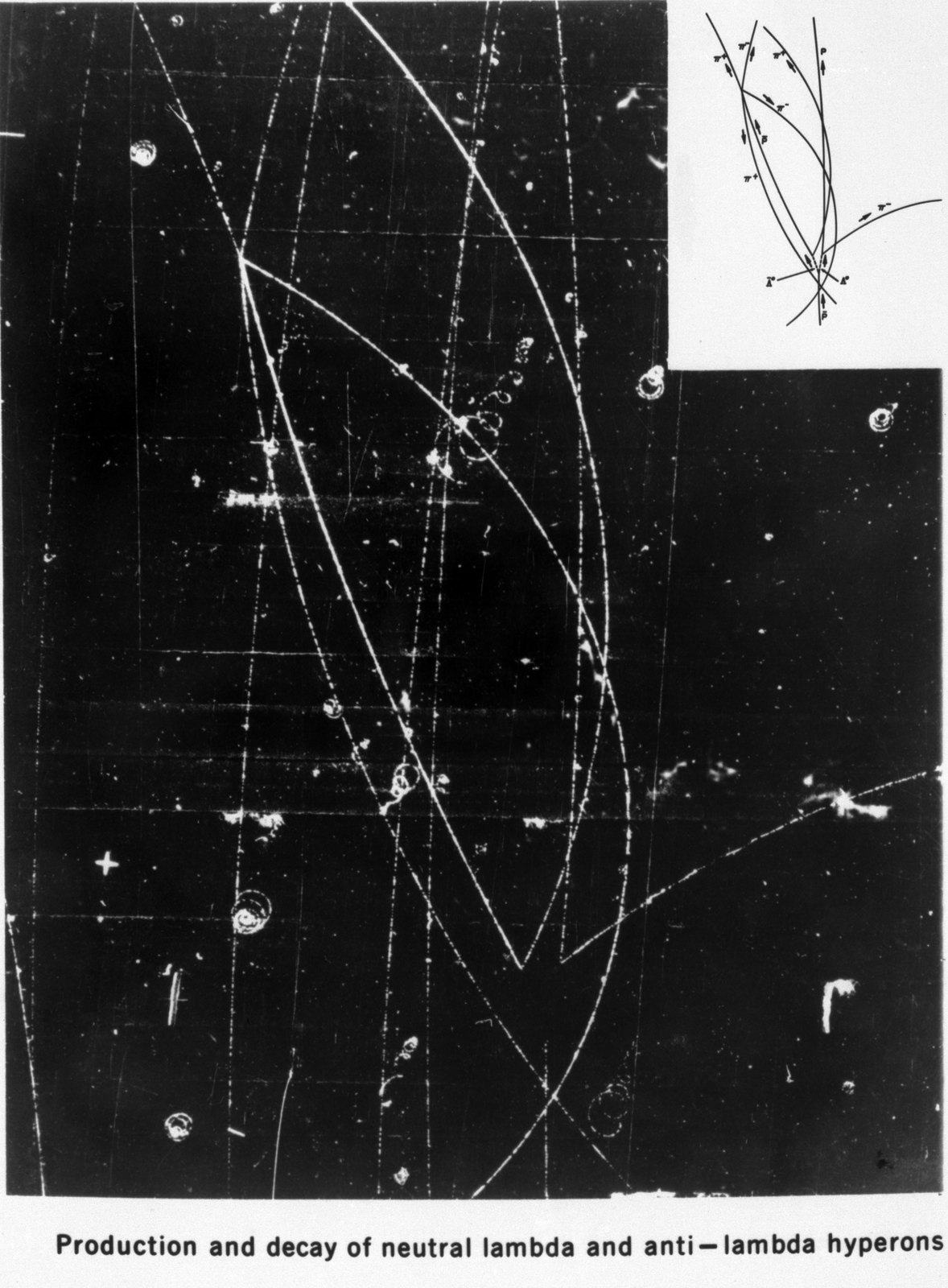
Production and decay of neutral lambda and anti-lambda hyperons. Patent release 10-5-1962. Photograph taken July 8, 1959. Bubble Chamber-172A - DPLA - 48d28c1e94d935715c697adf9a6554c8
27 Production and decay of neutral lambda and anti-lambda hyperons. Patent release 10-5-1962. Photograph taken July 8, 1959. Bubble Chamber-172A - DPLA - 48d28c1e94d935715c697adf9a6554c8
Department of Energy. Lawrence Berkeley National Laboratory. Public Affairs Department. Strategic Resources Office. Photography Services. 2012 · Public domain · sourceDownloadFull resolution (1876×2547) ↗
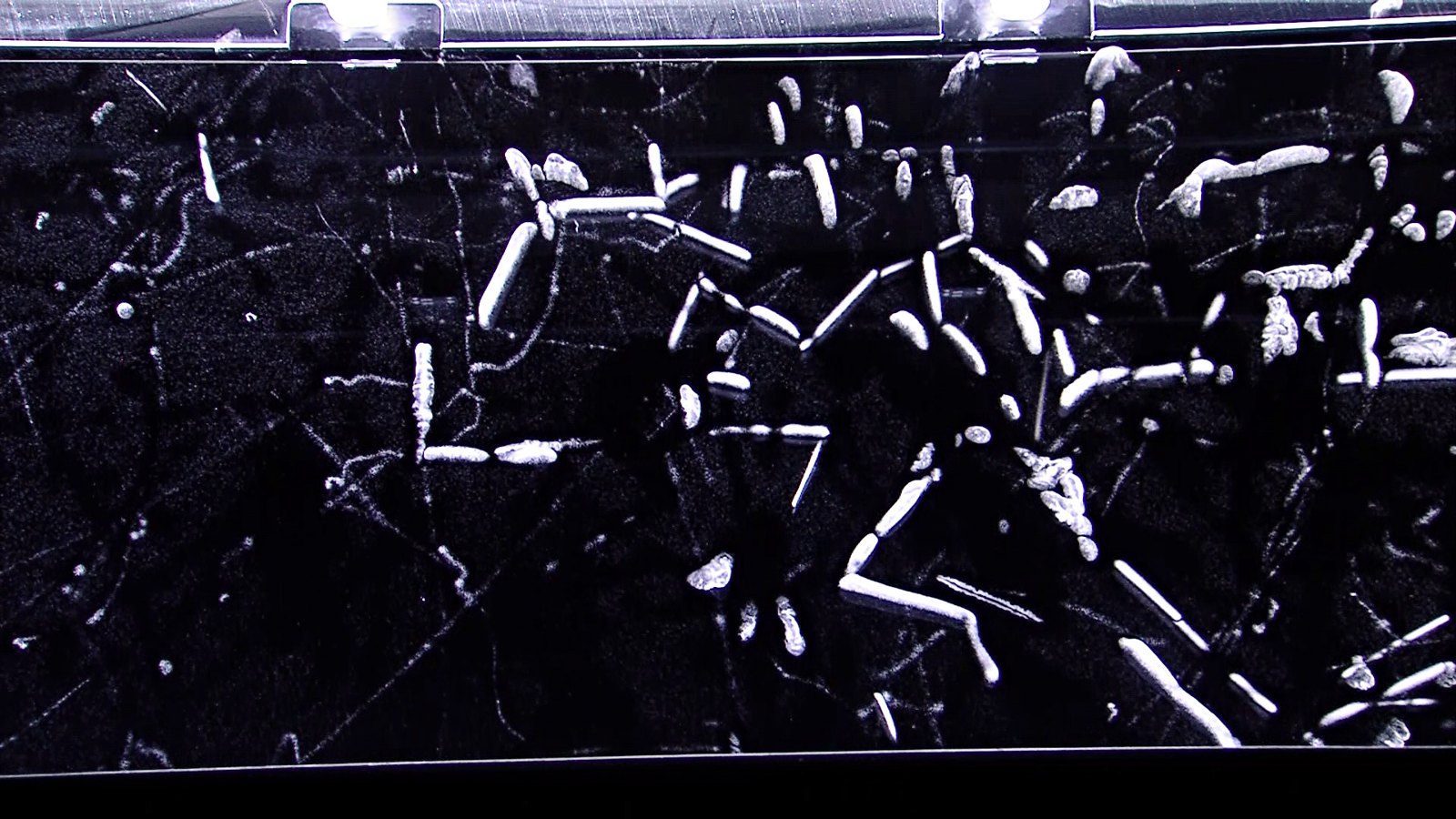
Alpha radiation in a cloud chamber
28 Alpha radiation in a cloud chamber
Nuledo · CC BY-SA 4.0 · sourceDownloadFull resolution (1920×1080) ↗
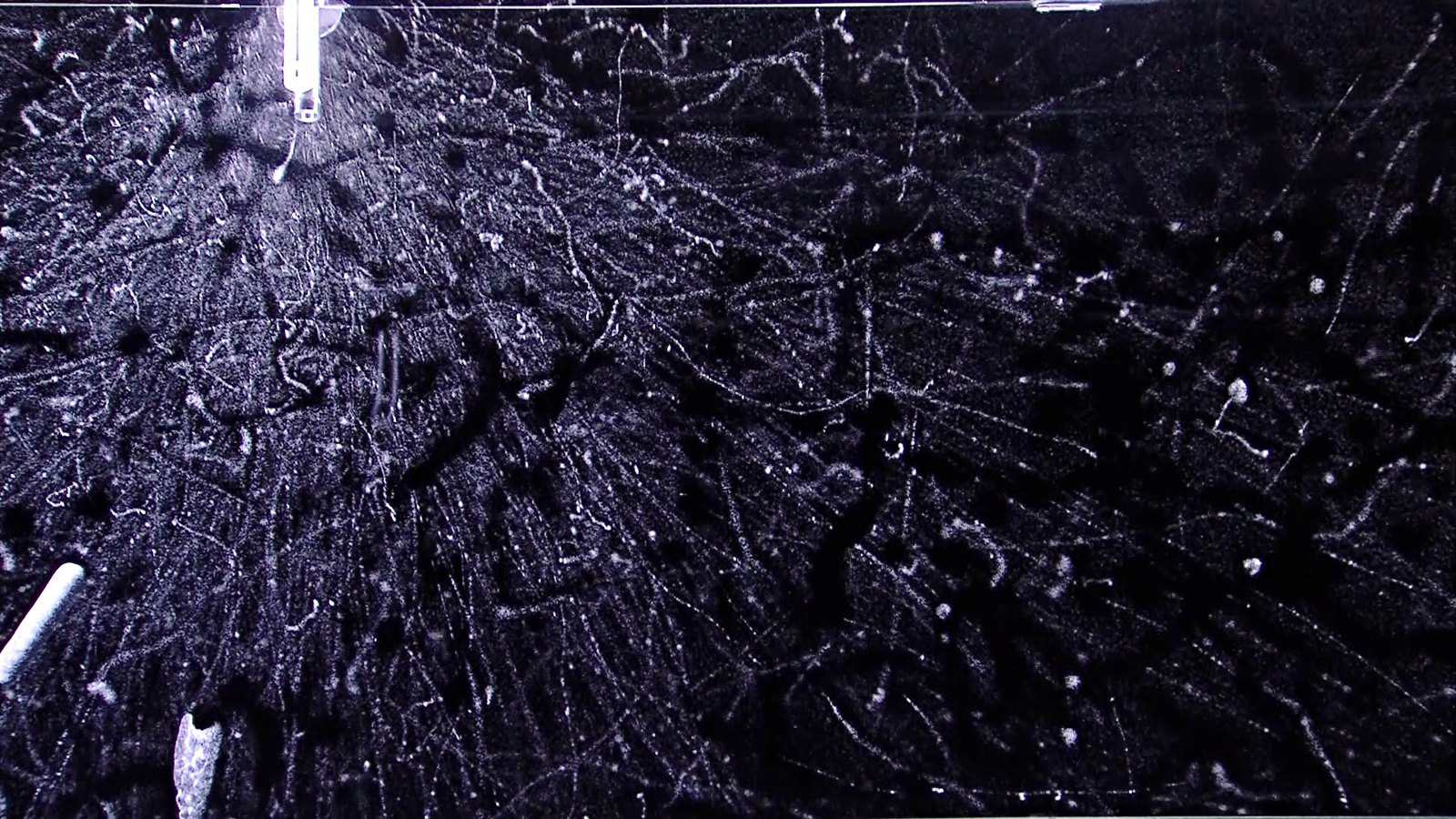
Beta radiation in a cloud chamber
29 Beta radiation in a cloud chamber
Nuledo · CC BY-SA 4.0 · sourceDownloadFull resolution (1920×1080) ↗
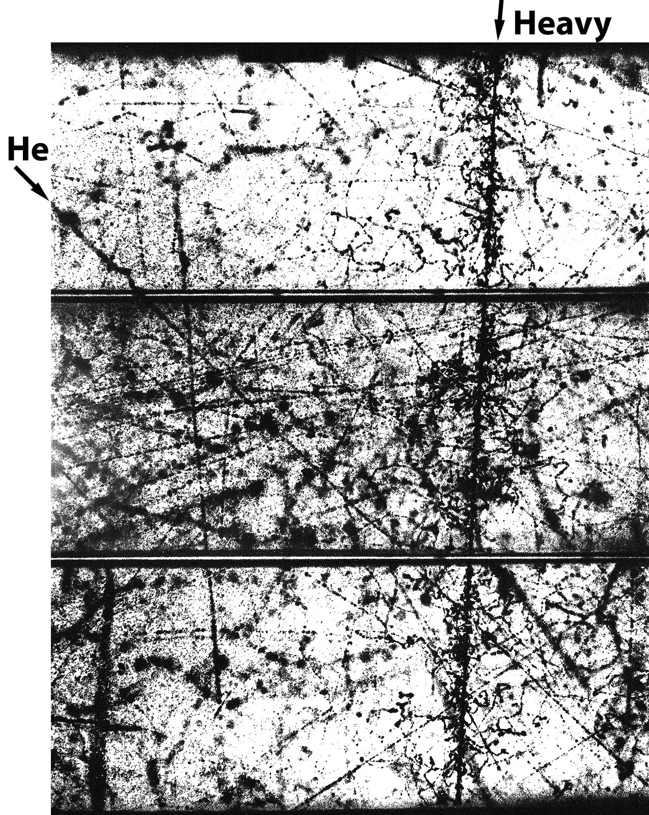
Cloud Chamber Photo of Heavy Nucleus
30 Cloud Chamber Photo of Heavy Nucleus
James A. Earl · CC BY 3.0 · sourceDownloadFull resolution (1530×1921) ↗
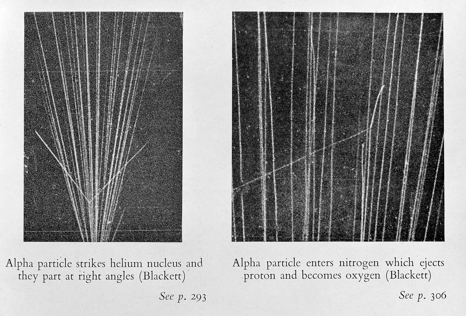
Experiments by Blackett in a Wilson Cloud Chamber. Wellcome M0015316
31 Experiments by Blackett in a Wilson Cloud Chamber. Wellcome M0015316
Unknown · CC BY 4.0 · sourceDownloadFull resolution (3970×2687) ↗
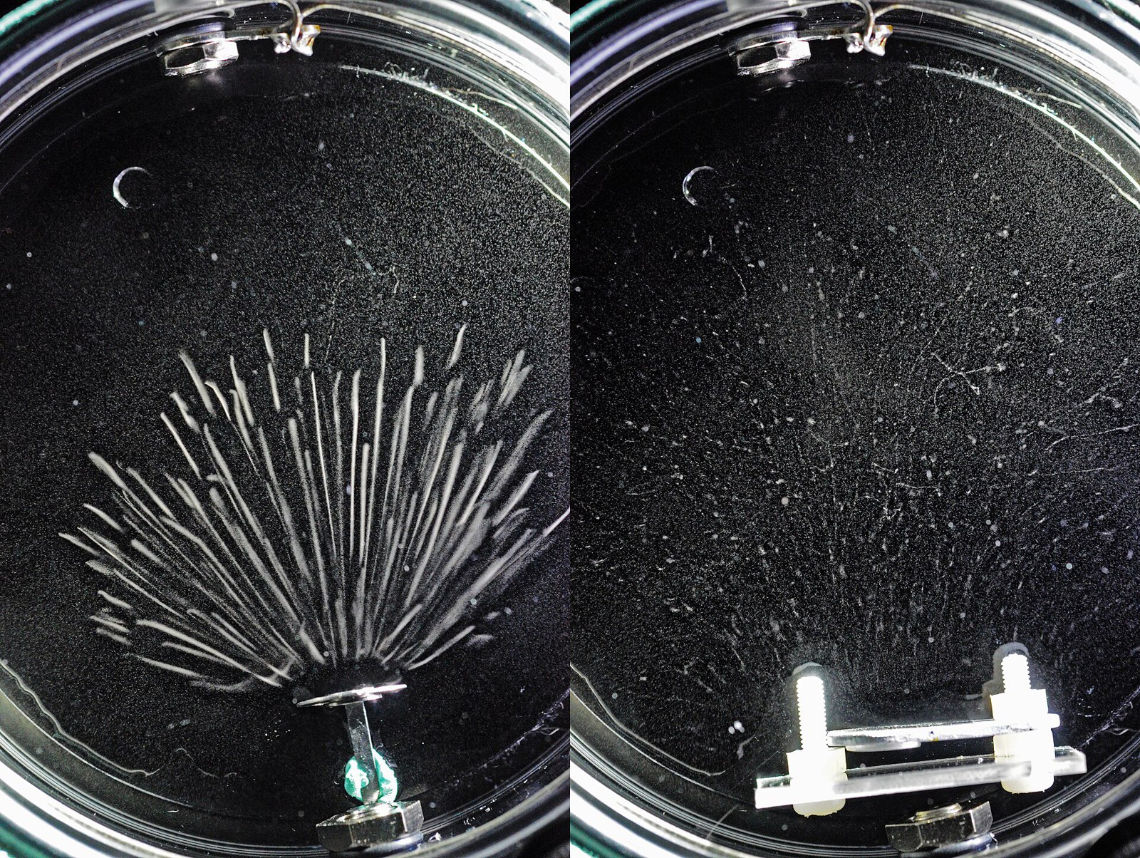
Particle Tracks in AWAN Expansion Cloud Chamber
32 Particle Tracks in AWAN Expansion Cloud Chamber
Kebuk awan · CC BY-SA 4.0 · sourceDownloadFull resolution (5696×4288) ↗
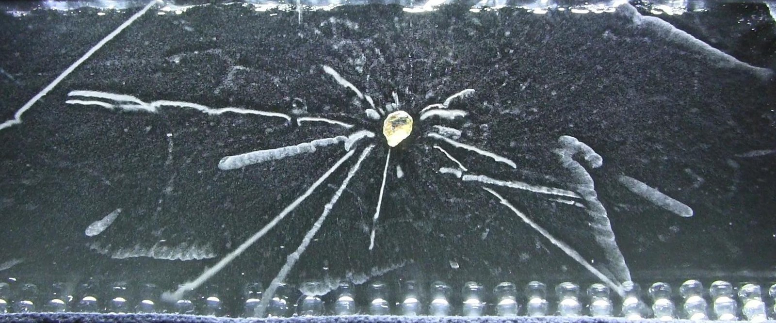
Radioactivity of a Thorite mineral seen in a cloud chamber
33 Radioactivity of a Thorite mineral seen in a cloud chamber
Cloudylabs · CC BY-SA 3.0 · sourceDownloadFull resolution (1816×756) ↗
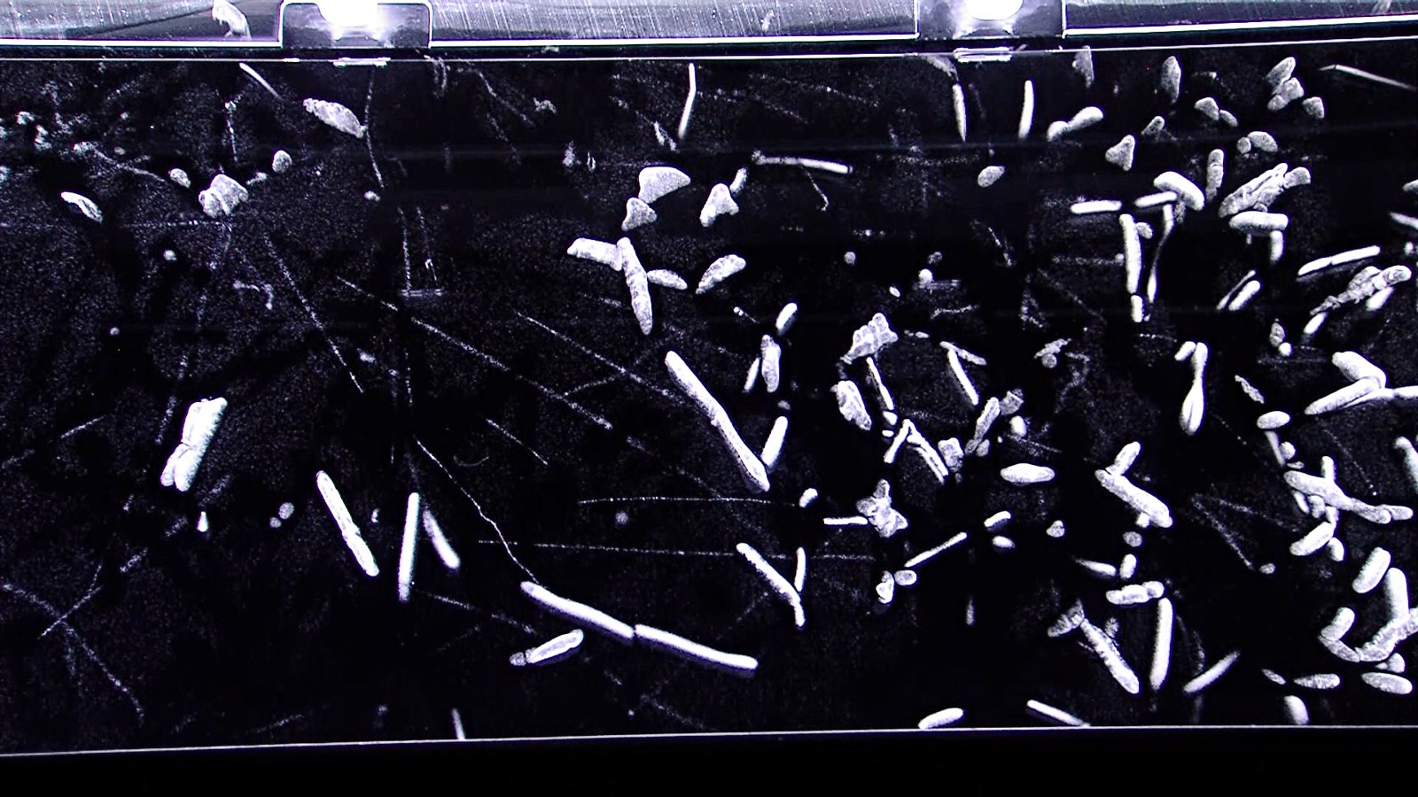
Radon decay in a cloud chamber
34 Radon decay in a cloud chamber
Nuledo · CC BY-SA 4.0 · sourceDownloadFull resolution (1920×1080) ↗
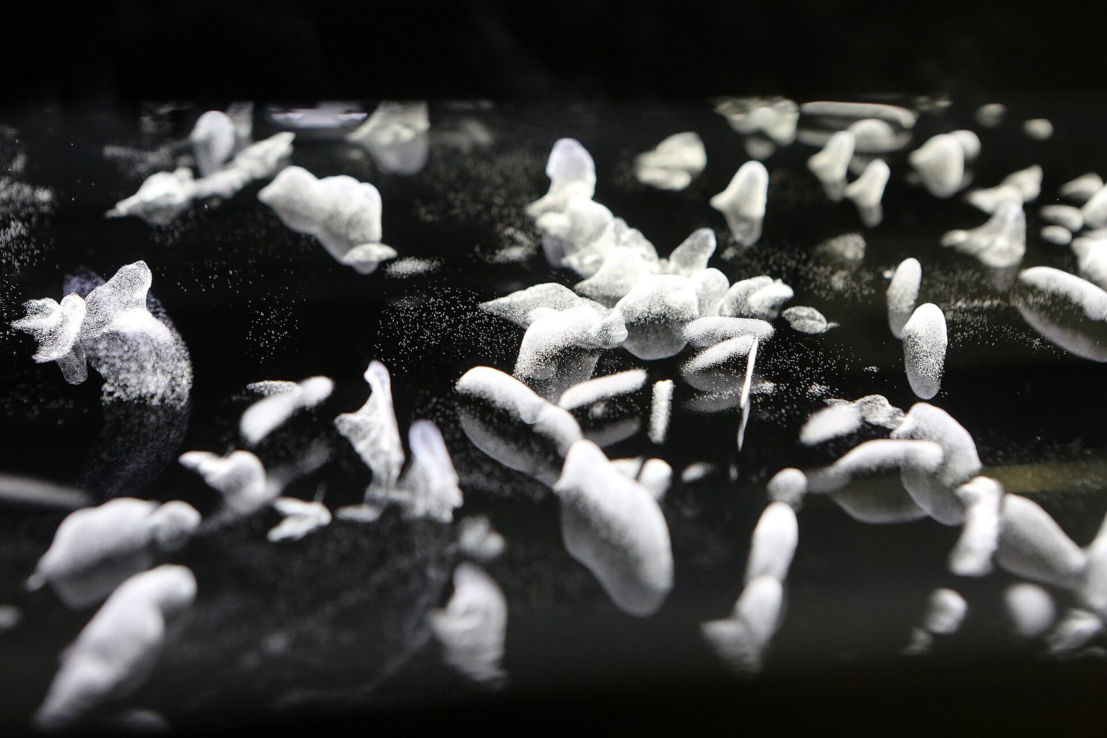
Vapor being used in a cloud chamber
35 Vapor being used in a cloud chamber
Nuledo · CC BY-SA 4.0 · sourceDownloadFull resolution (5760×3840) ↗

Cells, micrographs & radiolaria
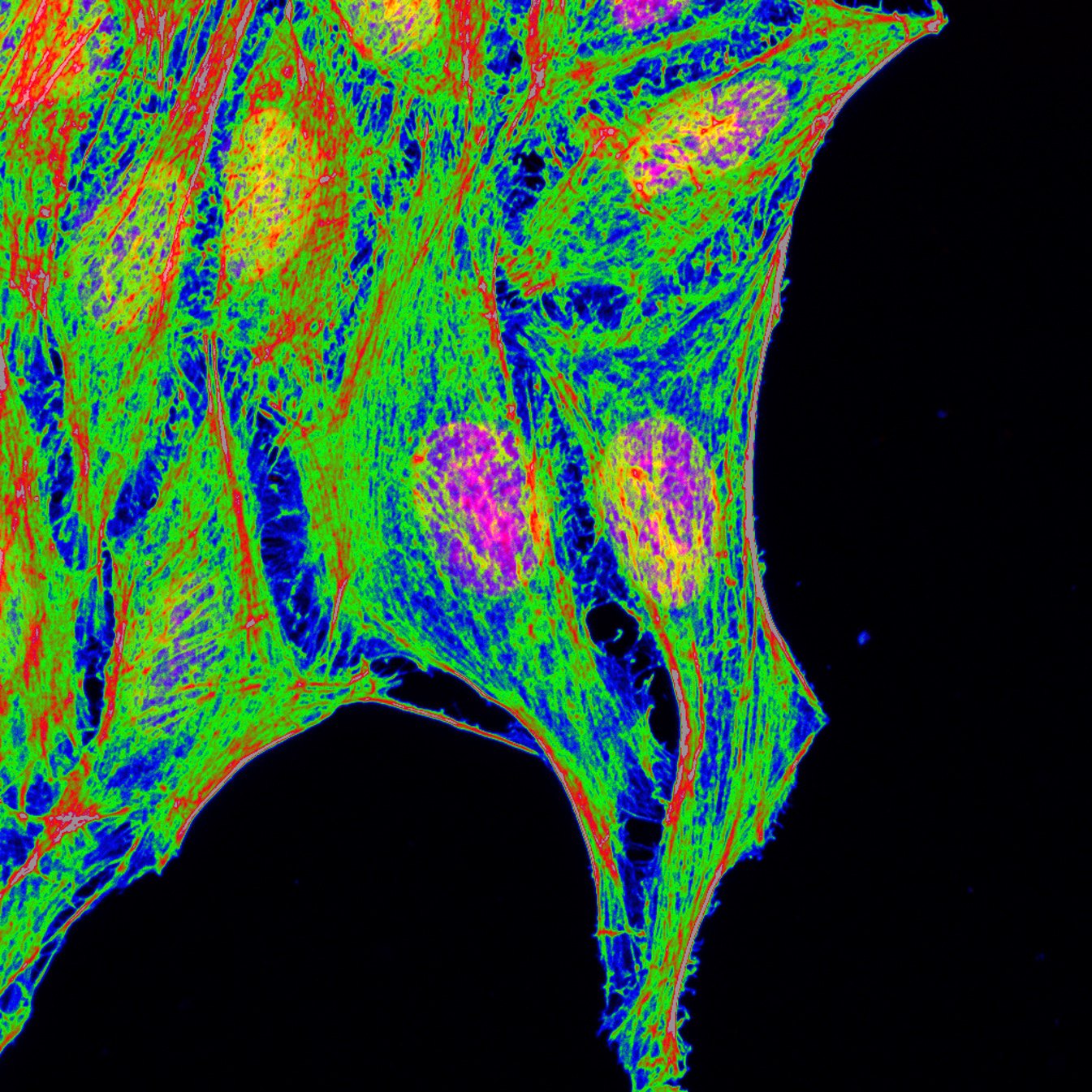
Confocal micrograph of stained HeLa
36 Confocal micrograph of stained HeLa
Dedyseptiadi7 · CC BY-SA 4.0 · sourceDownloadFull resolution (1345×1345) ↗
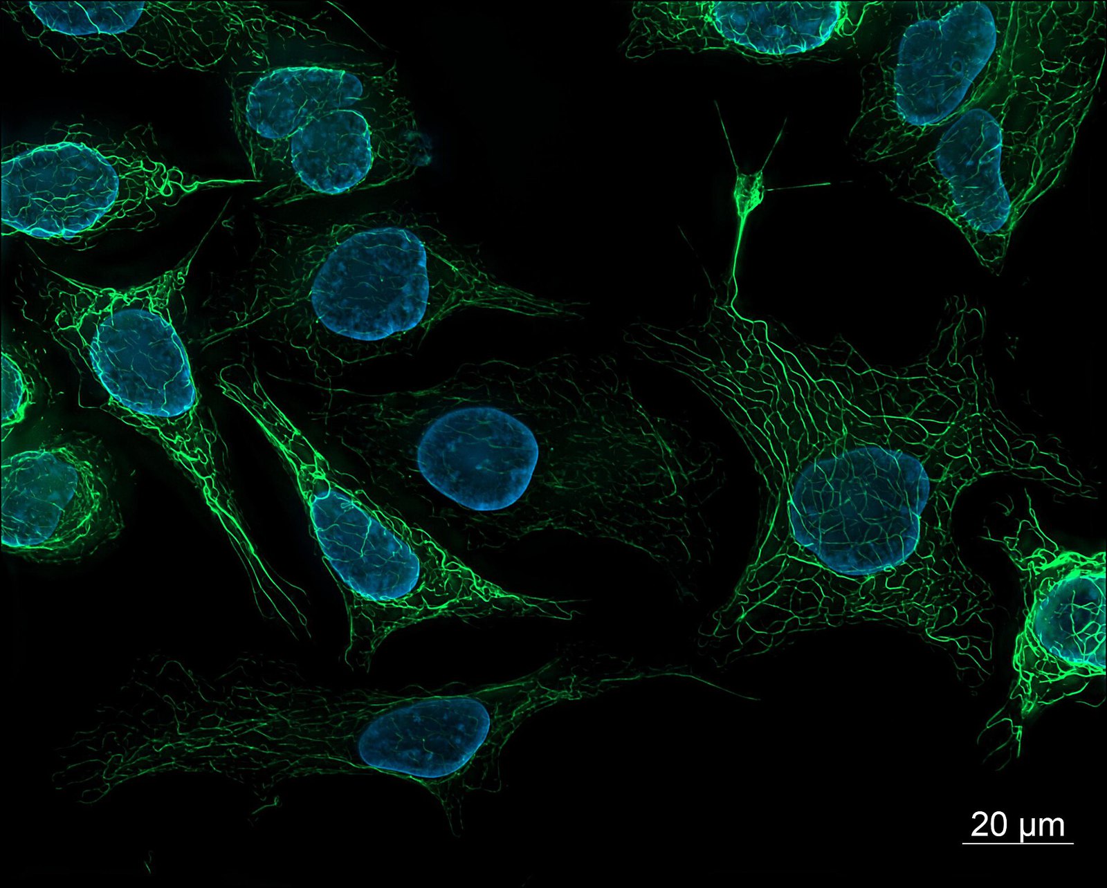
SK8-18-2 human derived cells, fluorescence microscopy (29942101073)
37 SK8-18-2 human derived cells, fluorescence microscopy (29942101073)
ZEISS Microscopy from Germany · CC BY 2.0 · sourceDownloadFull resolution (2758×2214) ↗
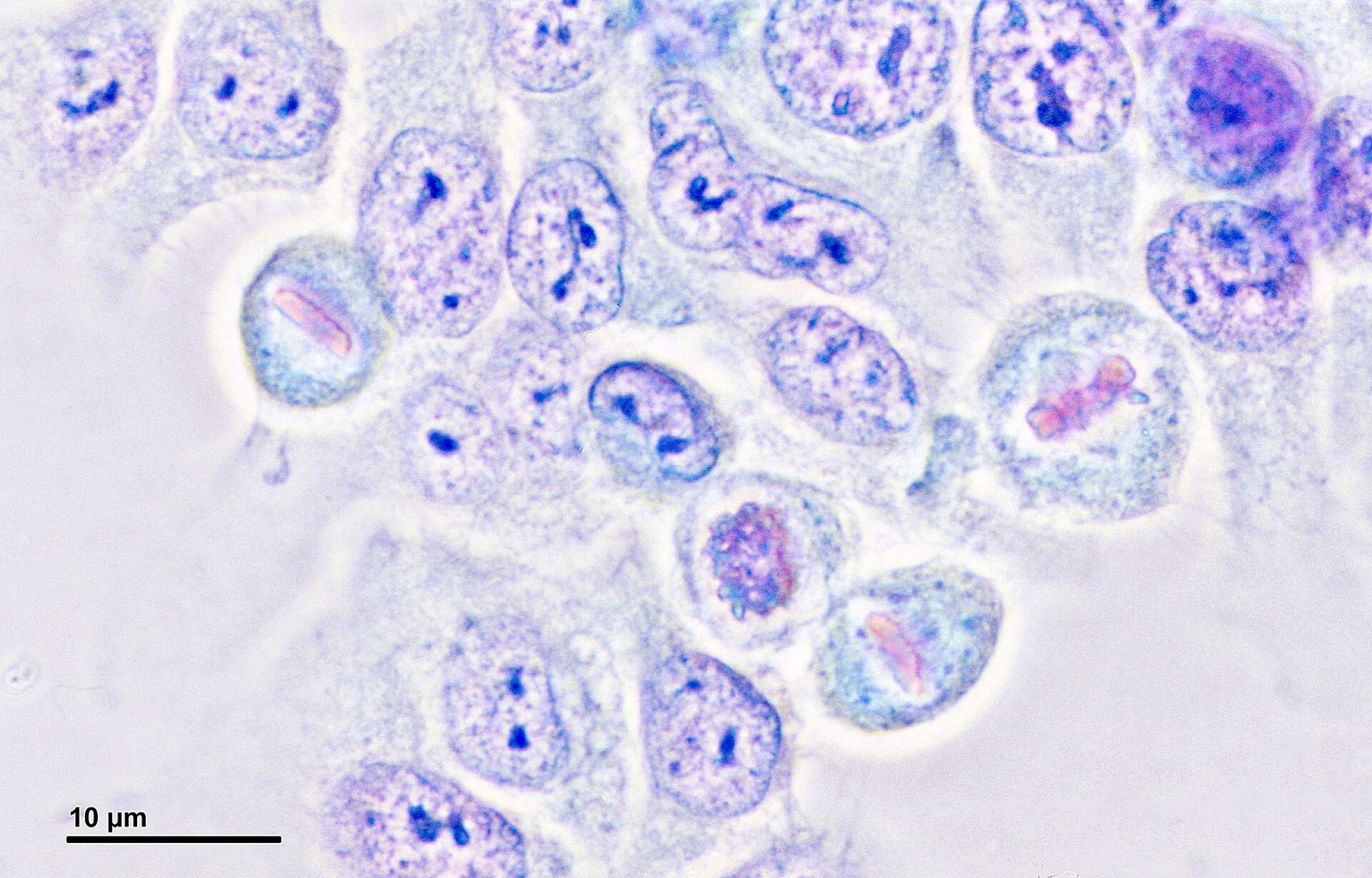
Cell culture (HeLa cells) (261 16) Cell culture (HeLa cells) - metaphase, prophase
38 Cell culture (HeLa cells) (261 16) Cell culture (HeLa cells) - metaphase, prophase
Doc. RNDr. Josef Reischig, CSc. · CC BY-SA 3.0 · sourceDownloadFull resolution (3751×2401) ↗
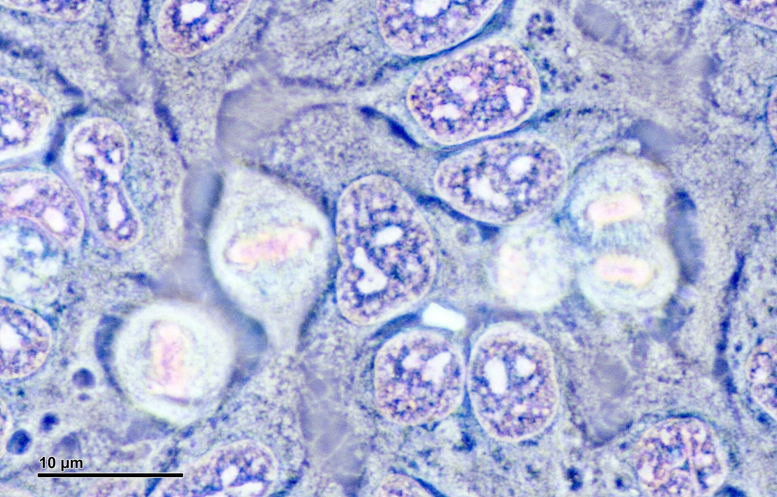
Cell culture (HeLa cells) (261 17) Cell culture (HeLa cells) - anaphase, metaphase
39 Cell culture (HeLa cells) (261 17) Cell culture (HeLa cells) - anaphase, metaphase
Doc. RNDr. Josef Reischig, CSc. · CC BY-SA 3.0 · sourceDownloadFull resolution (3751×2401) ↗
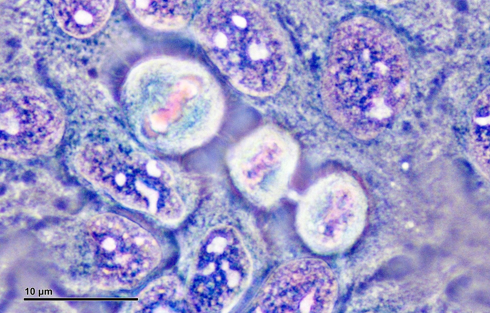
Cell culture (HeLa cells) (261 18) Cell culture (HeLa cells) - metaphase, telophase
40 Cell culture (HeLa cells) (261 18) Cell culture (HeLa cells) - metaphase, telophase
Doc. RNDr. Josef Reischig, CSc. · CC BY-SA 3.0 · sourceDownloadFull resolution (3751×2401) ↗
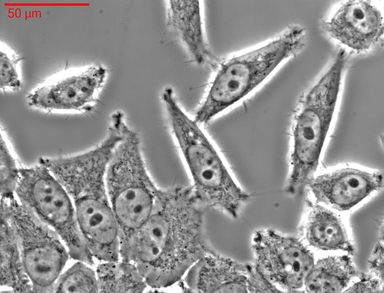
HELA-Zellen
41 HELA-Zellen
Fraunhofer-Institut für Biomedizinische Technik, St. Ingbert, Paul Anastasiadis, Eike Weiß · CC BY-SA 3.0 · sourceDownloadFull resolution (1344×1024) ↗
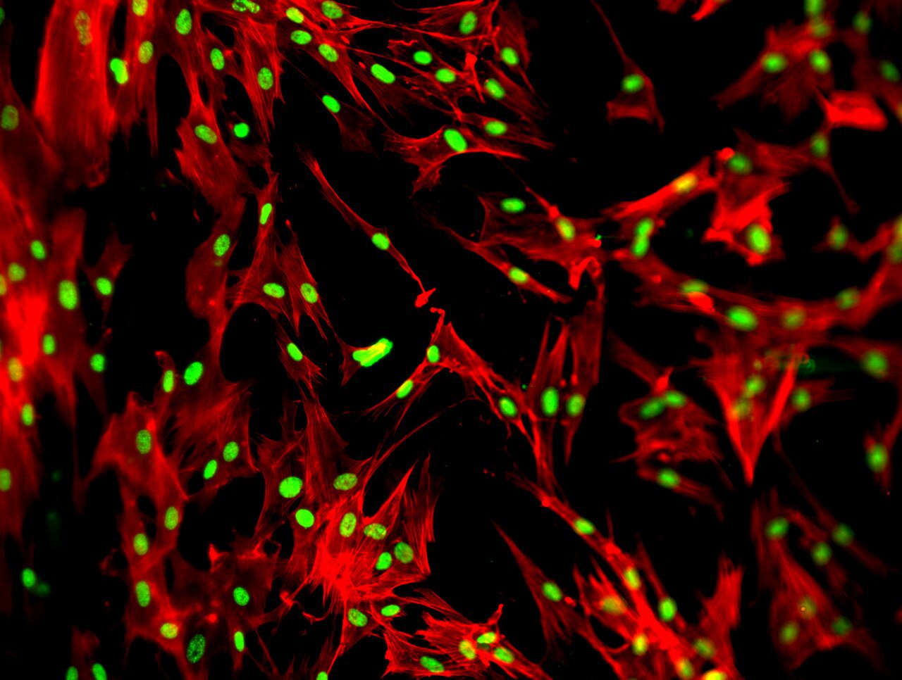
HELA cells stained with DAPI and Phalloidin
42 HELA cells stained with DAPI and Phalloidin
Arnatbio · CC BY-SA 4.0 · sourceDownloadFull resolution (1376×1038) ↗
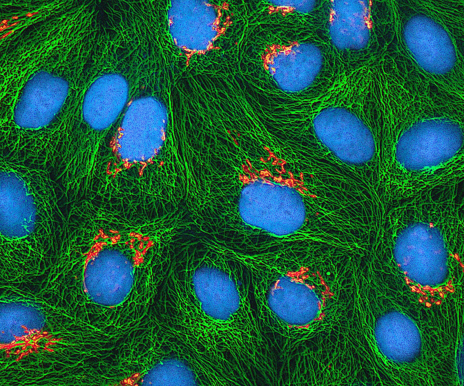
HeLa-I
43 HeLa-I
National Institutes of Health (NIH) · Public domain · sourceDownloadFull resolution (2400×1999) ↗
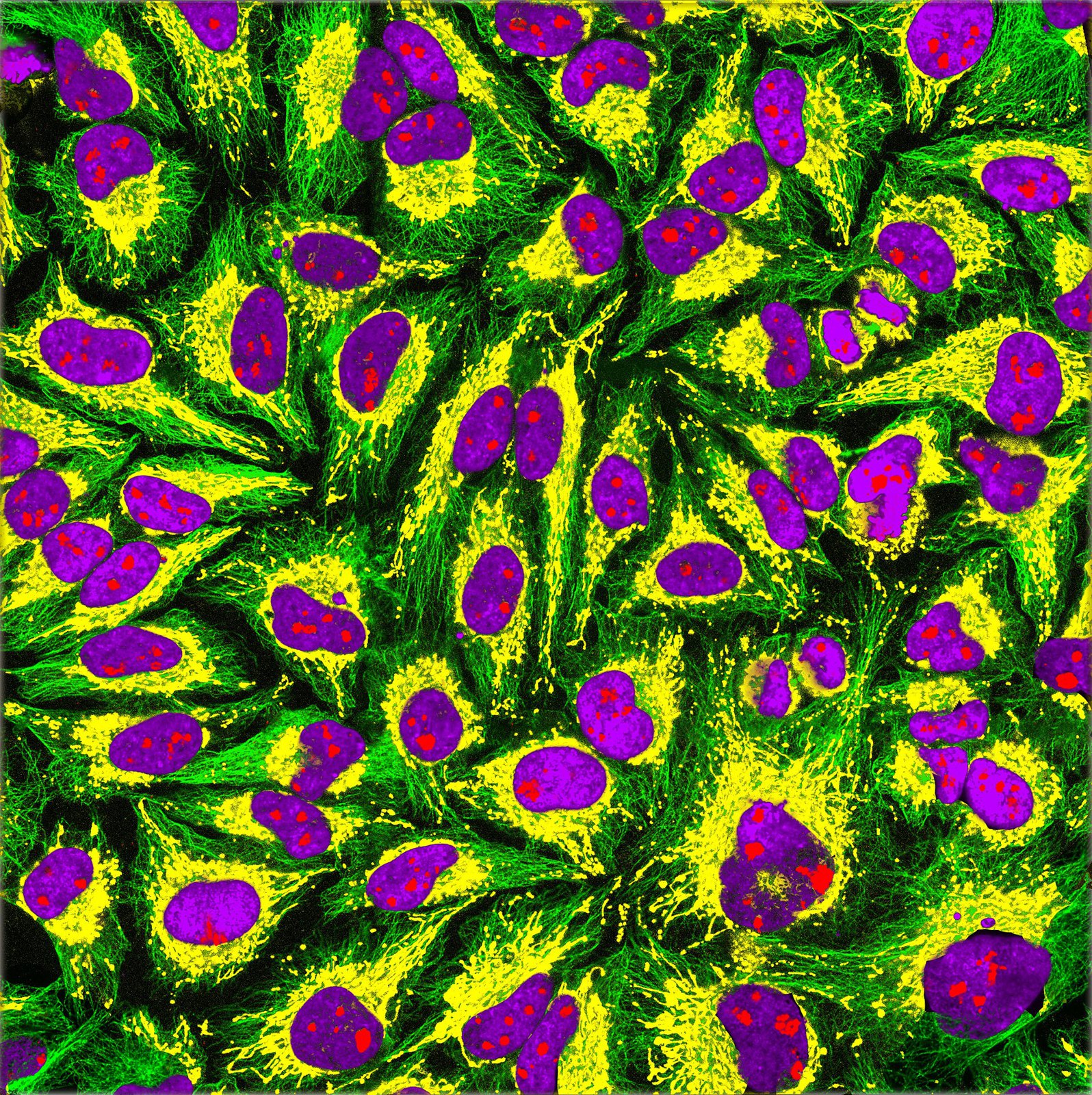
HeLa-Tubulin-HSP60-Fibrillarin-DNA
44 HeLa-Tubulin-HSP60-Fibrillarin-DNA
GerryShaw · CC BY-SA 4.0 · sourceDownloadFull resolution (2968×2976) ↗
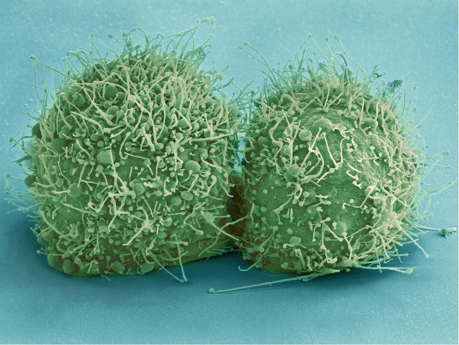
HeLa-V
45 HeLa-V
National Institutes of Health (NIH) · Public domain · sourceDownloadFull resolution (3200×2409) ↗
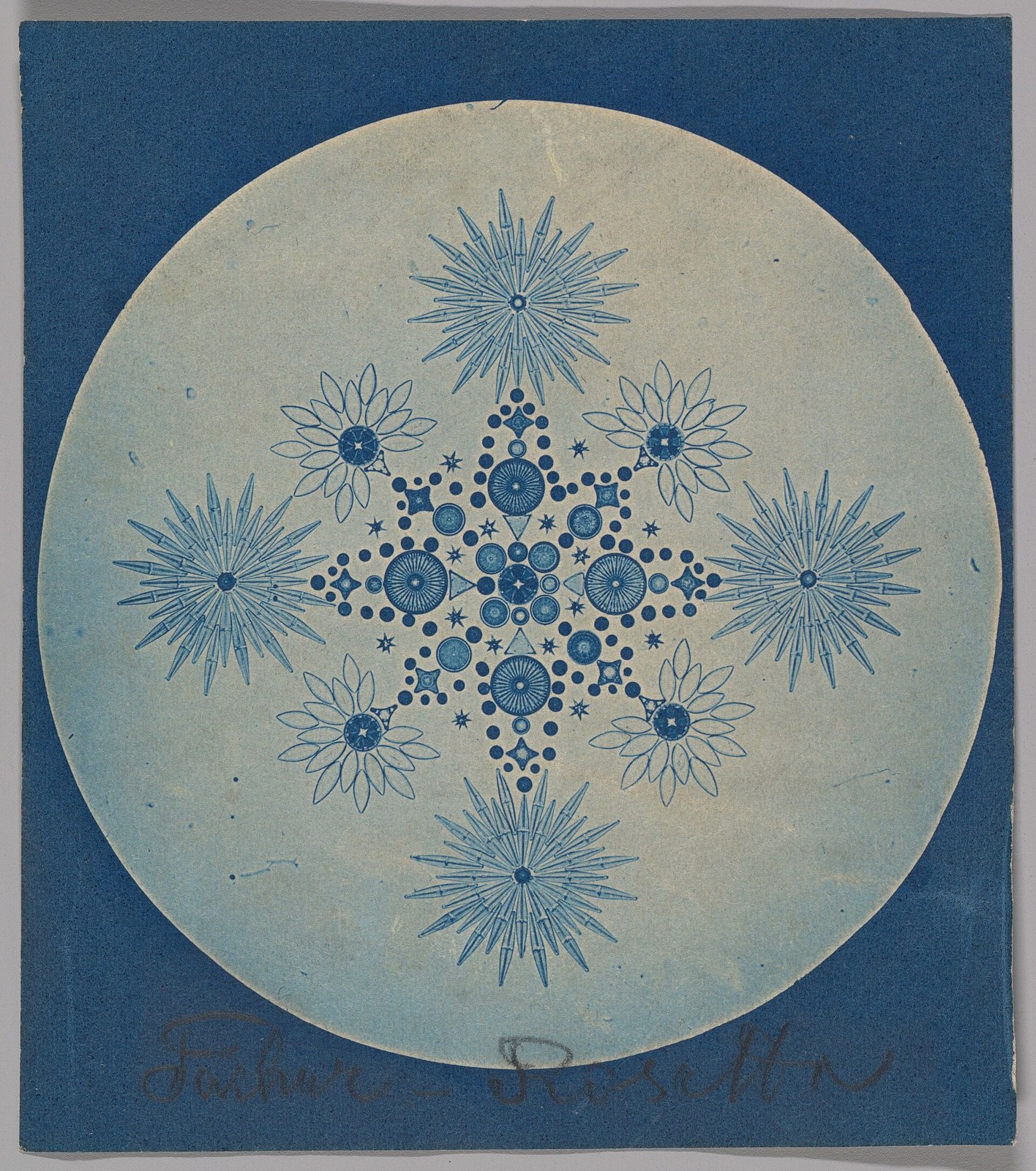
-Frustules of Diatoms- MET DP262066
46 -Frustules of Diatoms- MET DP262066
Creator:Julius Wiesner · CC0 · sourceDownloadFull resolution (2561×2893) ↗
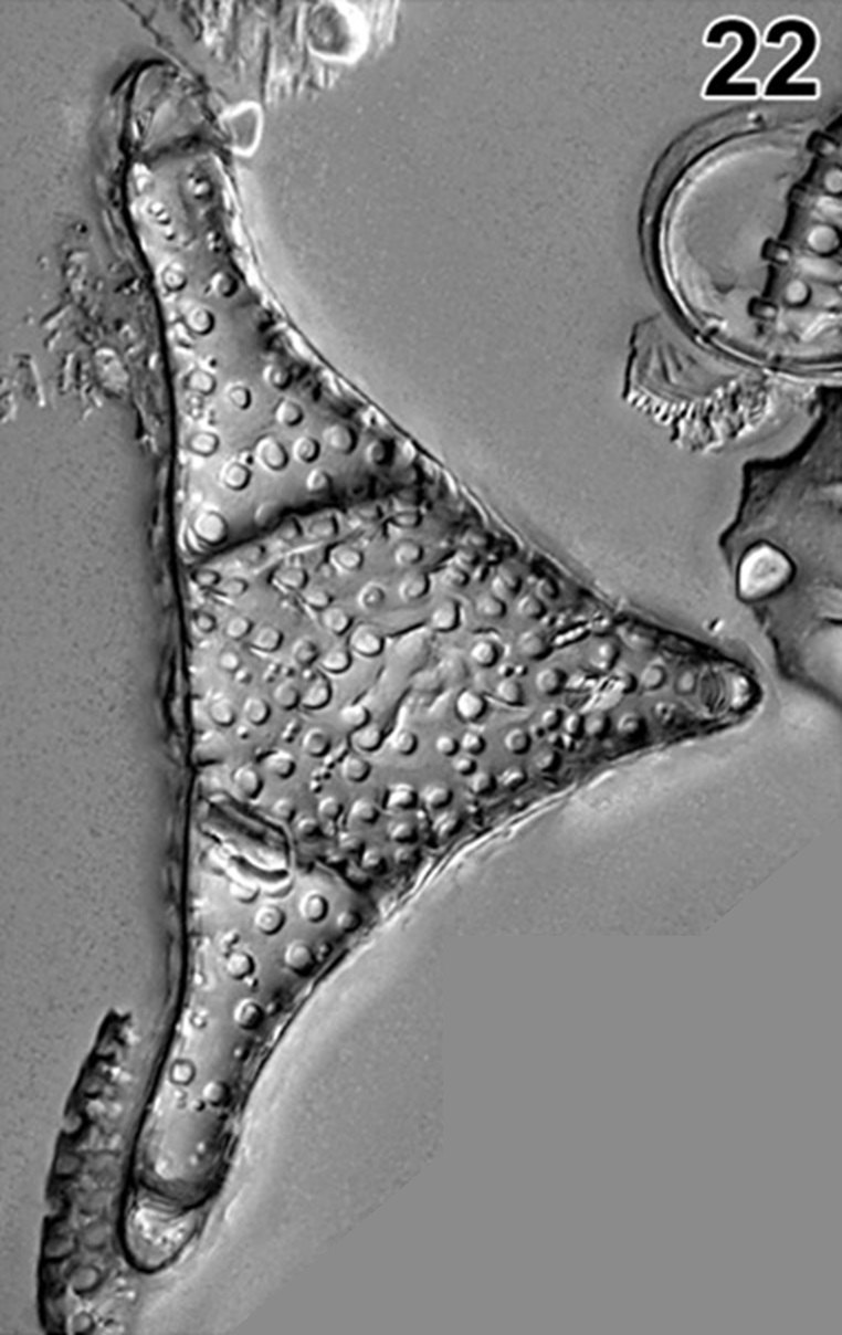
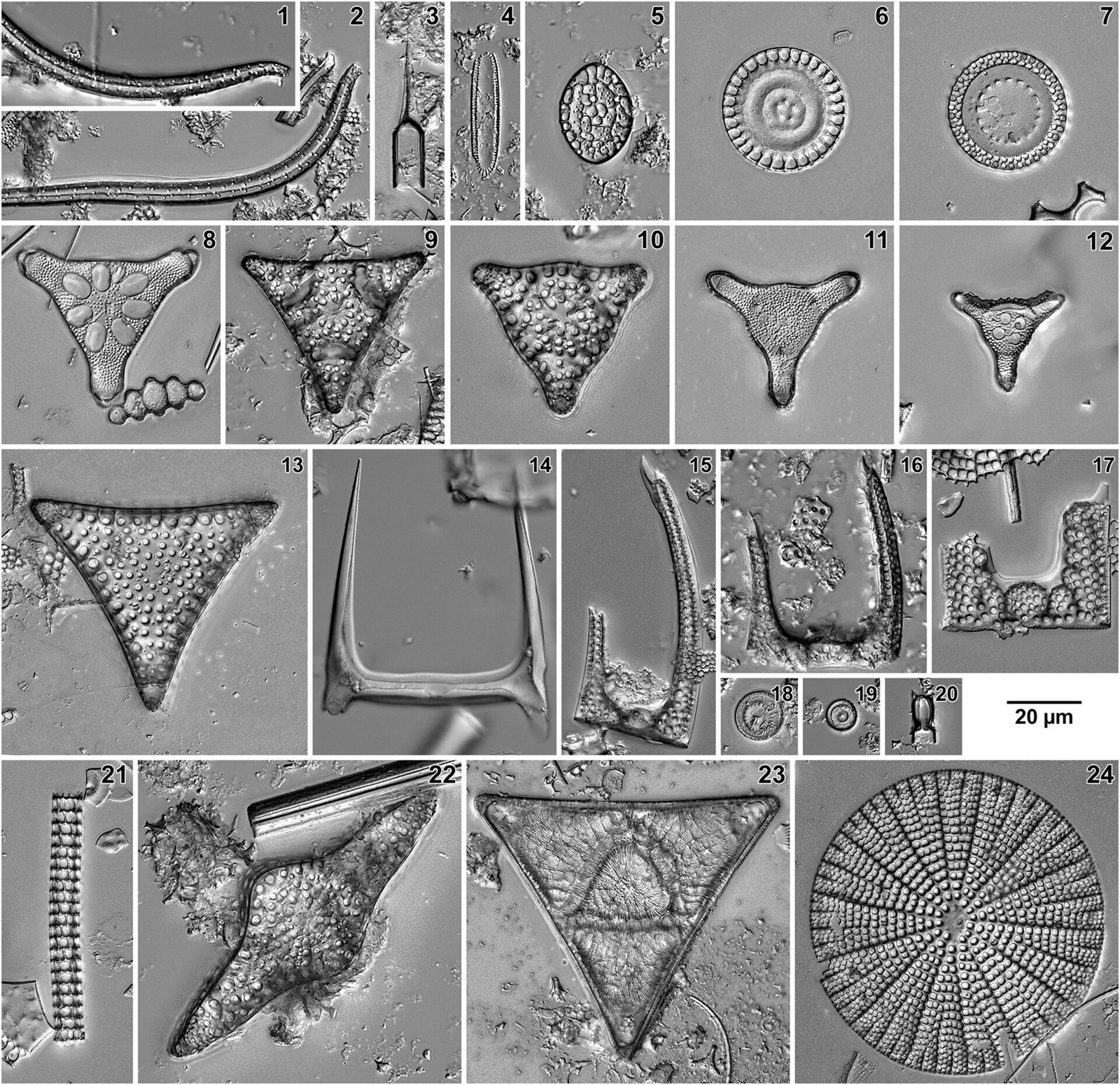
1-s2.0-S0025322720300566-pl1-fig22
47 1-s2.0-S0025322720300566-pl1-fig22
Jakub Witkowski, David M. Harwood, Bridget S. Wade, Karolina Bryłka · CC BY 4.0 · sourceDownloadFull resolution (762×1208) ↗
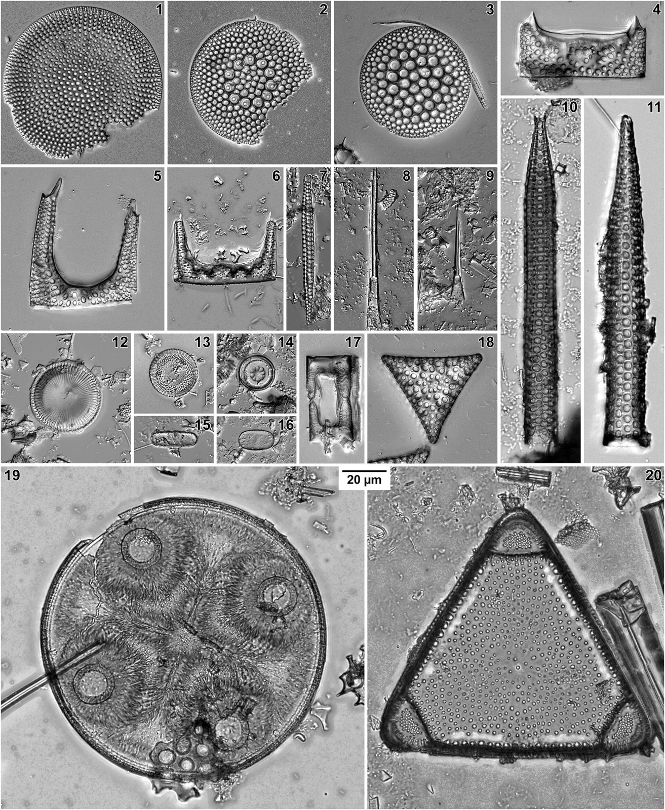
1-s2.0-S0025322720300566-pl2 lrg
48 1-s2.0-S0025322720300566-pl2 lrg
Jakub Witkowski, David M. Harwood, Bridget S. Wade, Karolina Bryłka · CC BY 4.0 · sourceDownloadFull resolution (2162×2634) ↗
1-s2.0-S0025322720300566-pl3 lrg
49 1-s2.0-S0025322720300566-pl3 lrg
Jakub Witkowski, David M. Harwood, Bridget S. Wade, Karolina Bryłka · CC BY 4.0 · sourceDownloadFull resolution (2162×2095) ↗
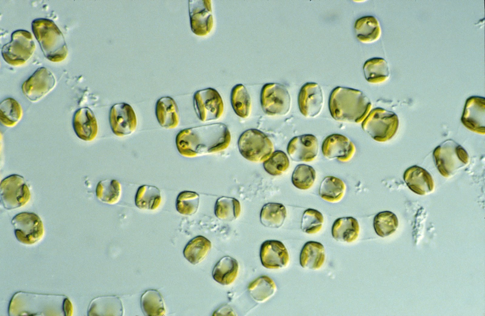
CSIRO ScienceImage 4905 diatom
50 CSIRO ScienceImage 4905 diatom
CSIRO, CSIRO · CC BY 3.0 · sourceDownloadFull resolution (1826×1191) ↗
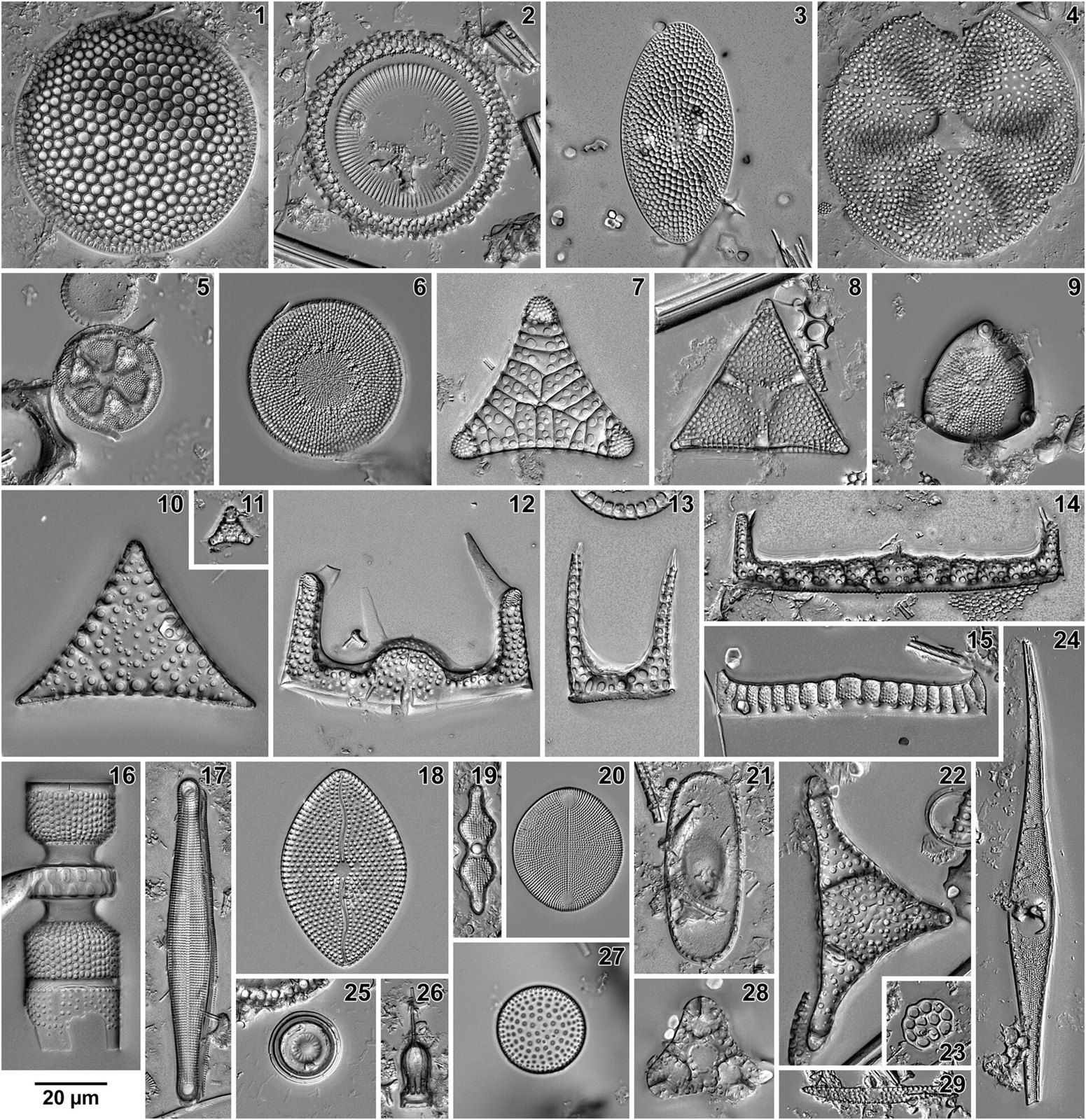
Eocene diatoms - Western North Atlantic
51 Eocene diatoms - Western North Atlantic
Jakub Witkowski, David M. Harwood, Bridget S. Wade, Karolina Bryłka · CC BY 4.0 · sourceDownloadFull resolution (2162×2231) ↗
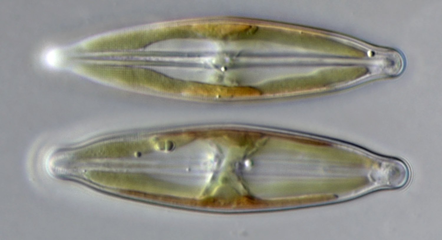
Naviculoid diatom
52 Naviculoid diatom
Djpmapfer · CC BY-SA 4.0 · sourceDownloadFull resolution (1500×814) ↗
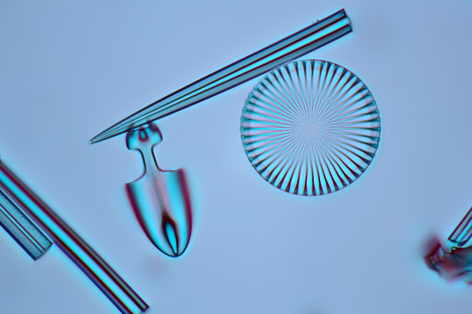
Rheinberg 2 diatom
53 Rheinberg 2 diatom
Frank Fox · CC BY-SA 3.0 de · sourceDownloadFull resolution (1920×1280) ↗
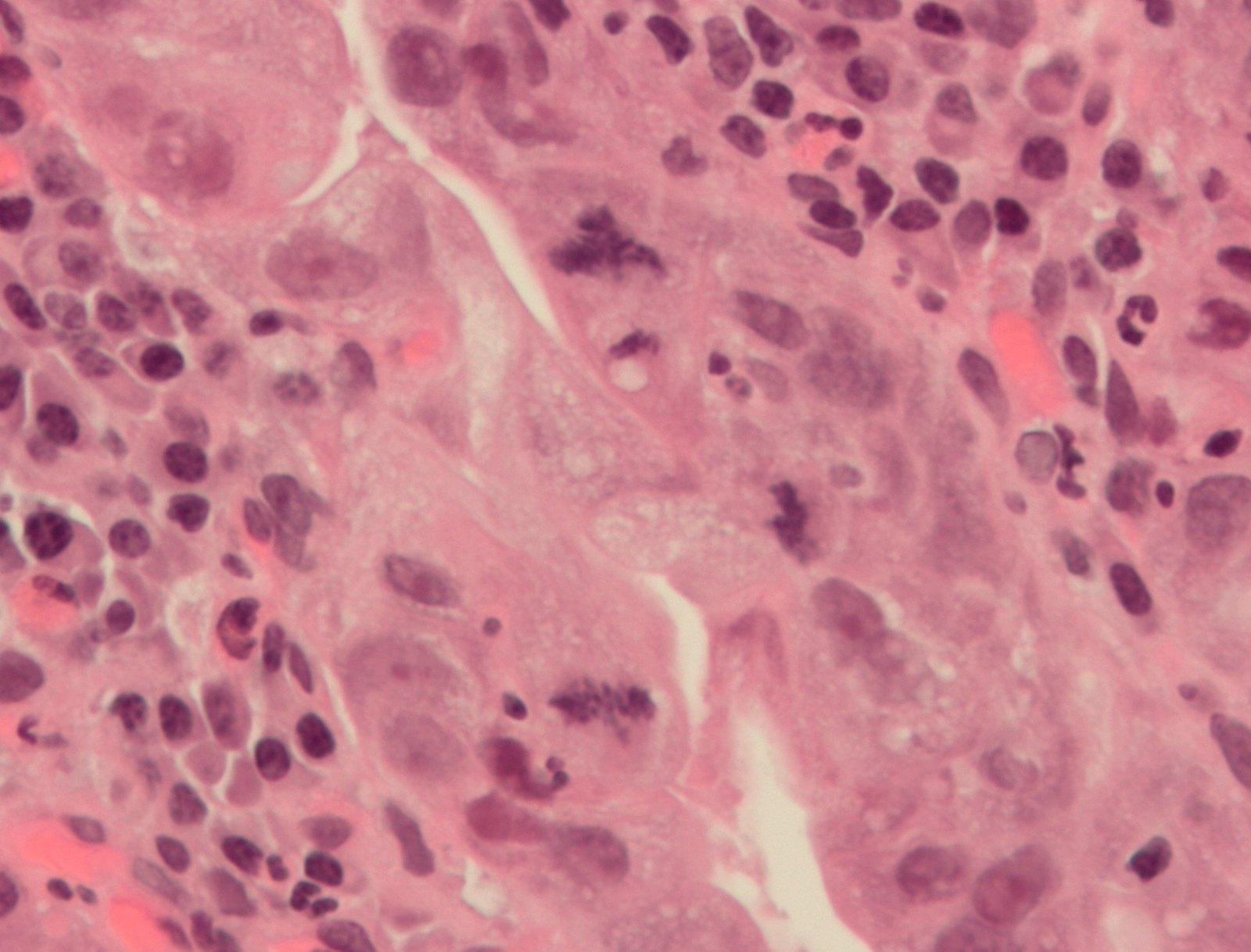
Atypical mitosis
54 Atypical mitosis
Nephron · CC BY-SA 3.0 · sourceDownloadFull resolution (1842×1402) ↗
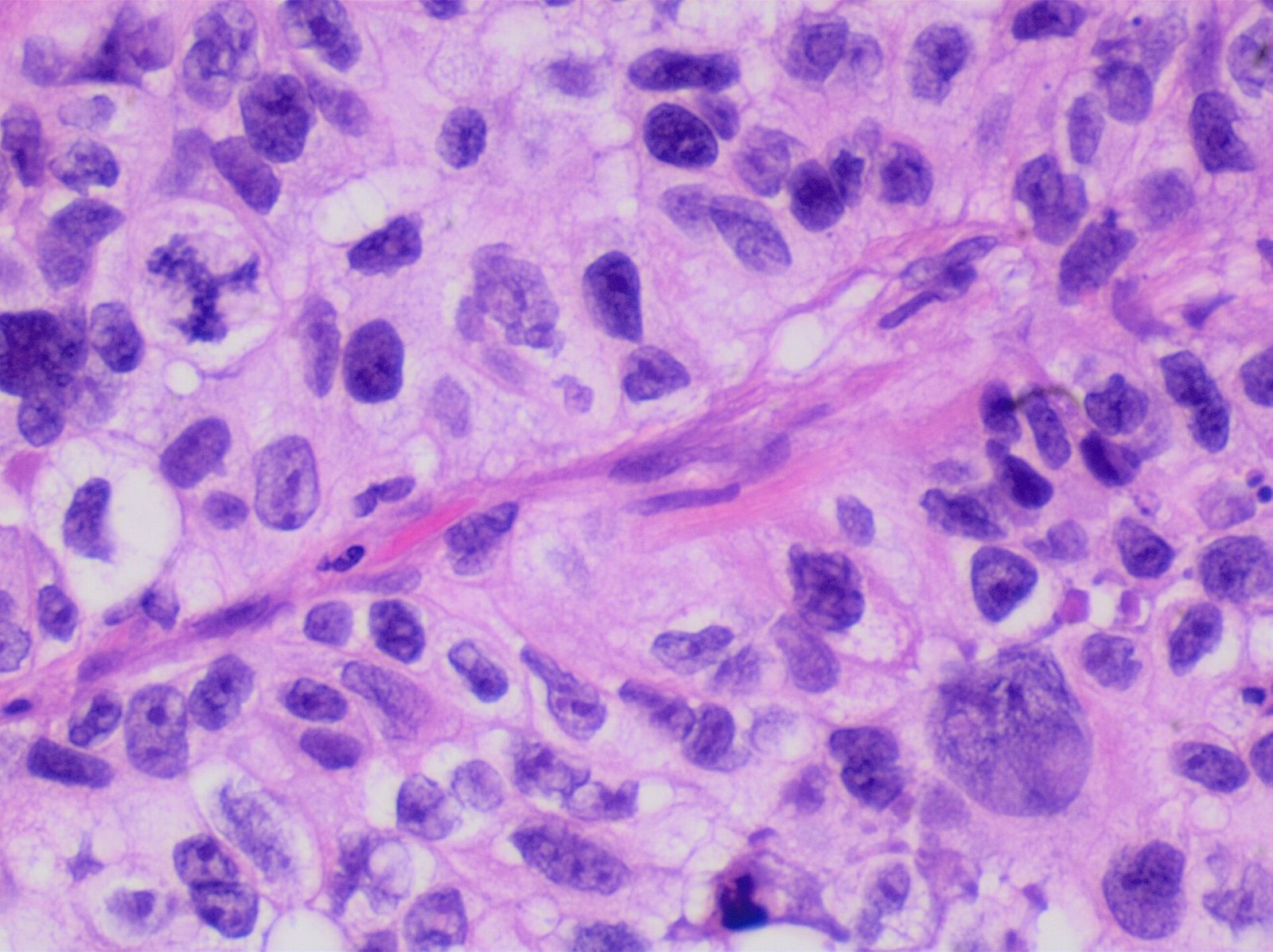
Histopathology of poorly differentiated lung adenocarcinoma
55 Histopathology of poorly differentiated lung adenocarcinoma
Mikael Häggström, M.D. Author info - Reusing images- Conflicts of interest: None Mikael Häggström, M.D. · CC0 · sourceDownloadFull resolution (2048×1532) ↗
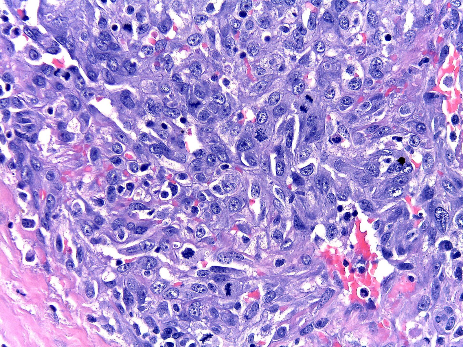
Kaposi sarcoma in a patient with HHV8-associated MCD
56 Kaposi sarcoma in a patient with HHV8-associated MCD
Amylacea · CC BY-SA 4.0 · sourceDownloadFull resolution (2048×1536) ↗
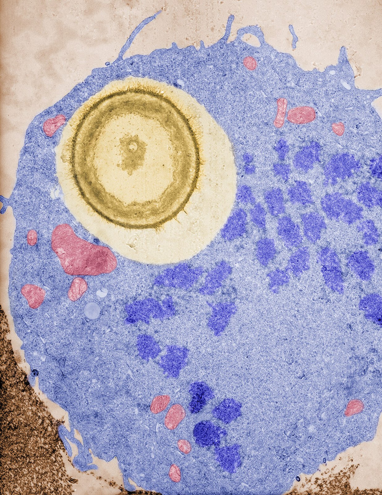
Macrophages undergo mitosis after ingesting a fungal cell
57 Macrophages undergo mitosis after ingesting a fungal cell
Carolina Coelho · CC BY-SA 4.0 · sourceDownloadFull resolution (2551×3300) ↗
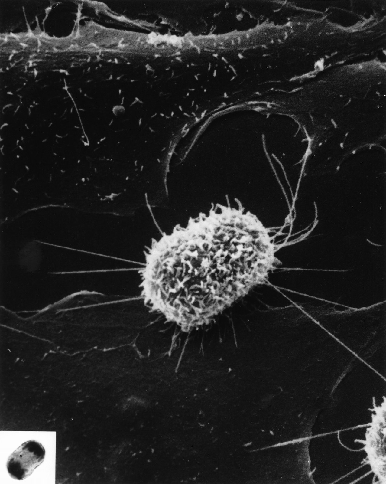
Mitosis (1)
58 Mitosis (1)
Unknown photographer · Public domain · sourceDownloadFull resolution (1800×2255) ↗
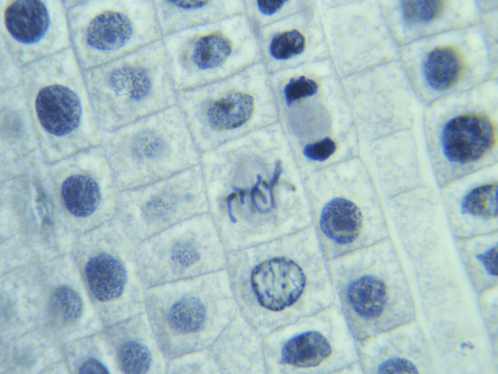
Mitosis in Onion Root - various stages (Metaphase and Telophase)
59 Mitosis in Onion Root - various stages (Metaphase and Telophase)
Natalierussell77 · CC BY-SA 4.0 · sourceDownloadFull resolution (4912×3684) ↗
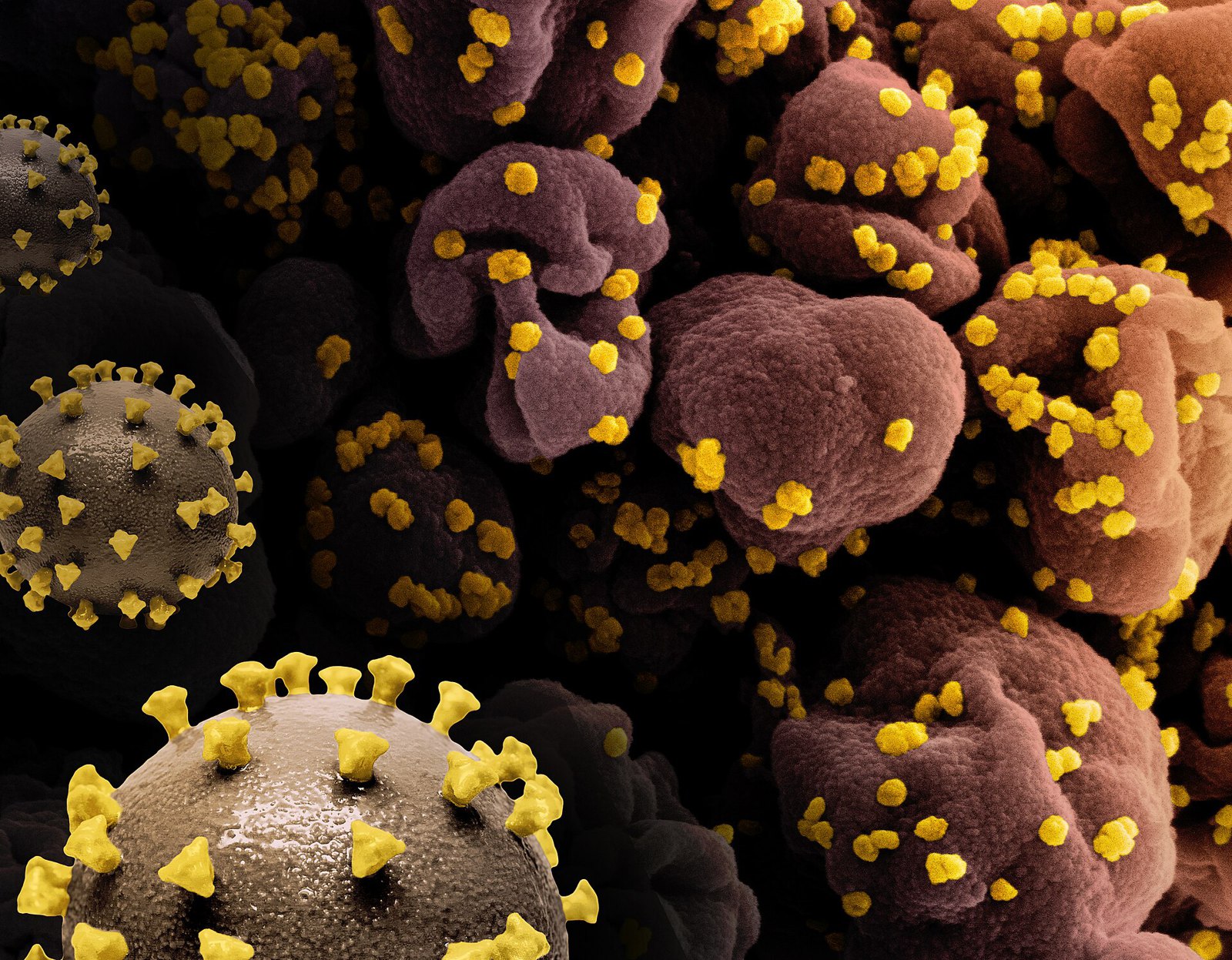
3D print models of SARS-CoV-2 virus (left, yellow spike proteins on a gray virus surface) and a colorized scanning electron micrograph of a cell infected with SARS-CoV-2 virus particles (yellow)
60 3D print models of SARS-CoV-2 virus (left, yellow spike proteins on a gray virus surface) and a colorized scanning electron micrograph of a cell infected with SARS-CoV-2 virus particles (yellow)
NIAID · CC BY 2.0 · sourceDownloadFull resolution (3207×2498) ↗
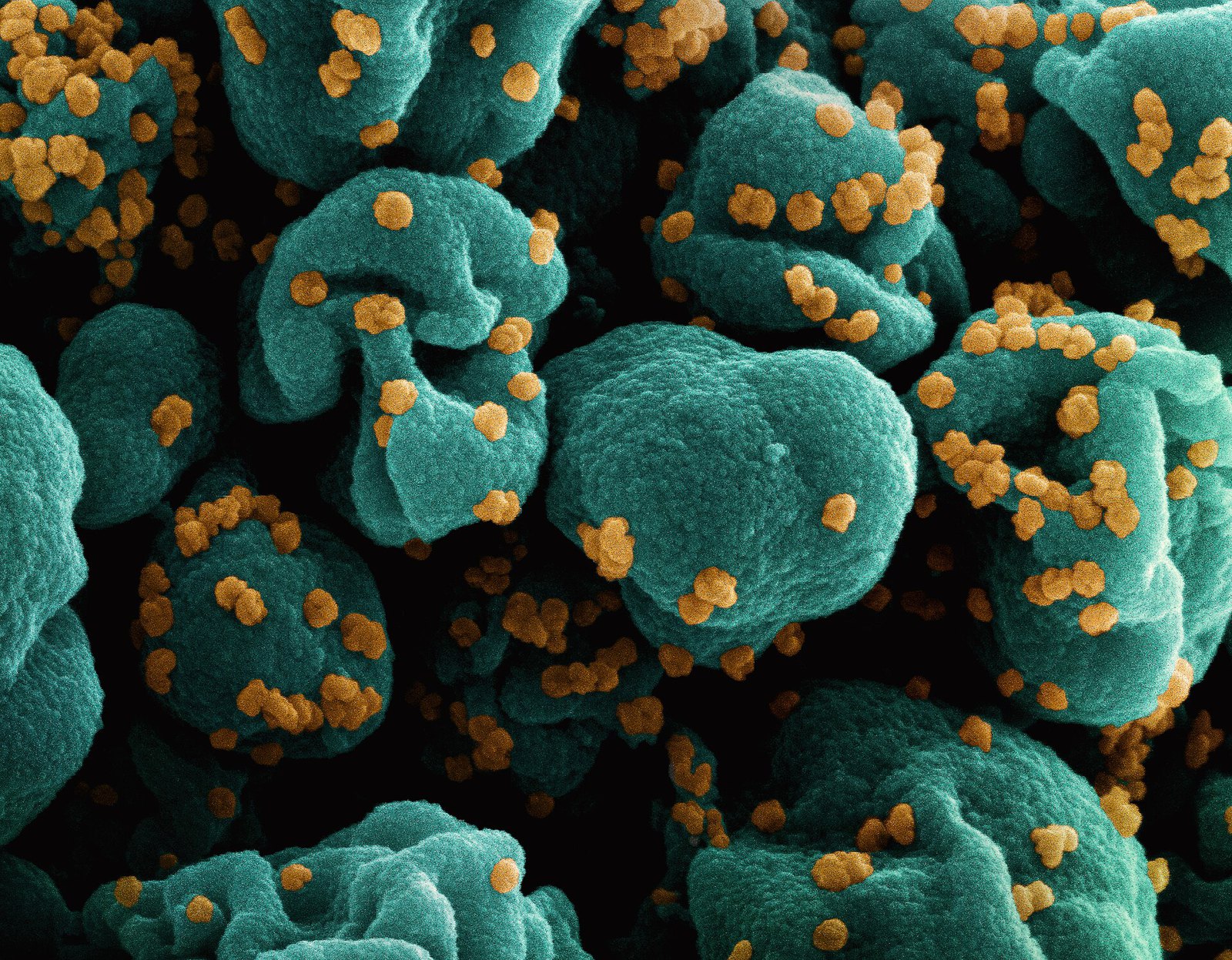
Colorized scanning electron micrograph of a cell (teal) infected with SARS-CoV-2 virus particles (orange), isolated from a patient sample
61 Colorized scanning electron micrograph of a cell (teal) infected with SARS-CoV-2 virus particles (orange), isolated from a patient sample
NIAID · CC BY 2.0 · sourceDownloadFull resolution (2719×2118) ↗
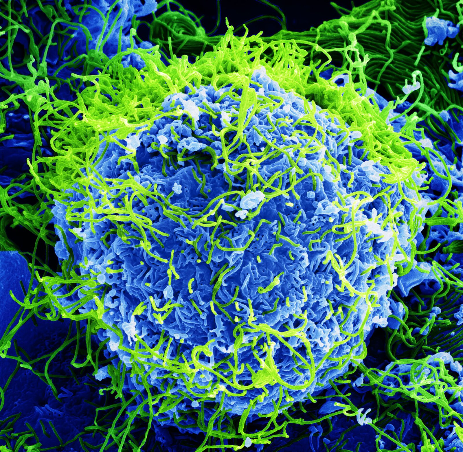
Ebola Virus - Electron Micrograph
62 Ebola Virus - Electron Micrograph
BernbaumJG · CC BY 4.0 · sourceDownloadFull resolution (3604×3518) ↗
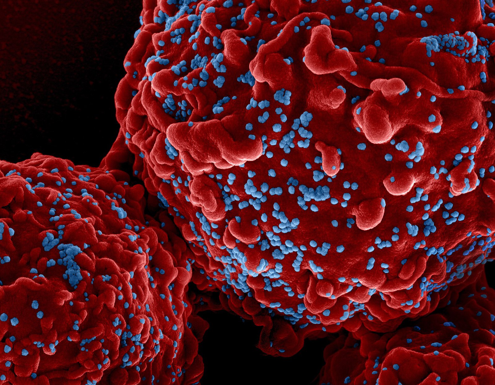
HIV-1 virus particles colorized scanning electron micrograph - 111
63 HIV-1 virus particles colorized scanning electron micrograph - 111
NIAID · CC BY 2.0 · sourceDownloadFull resolution (3406×2648) ↗
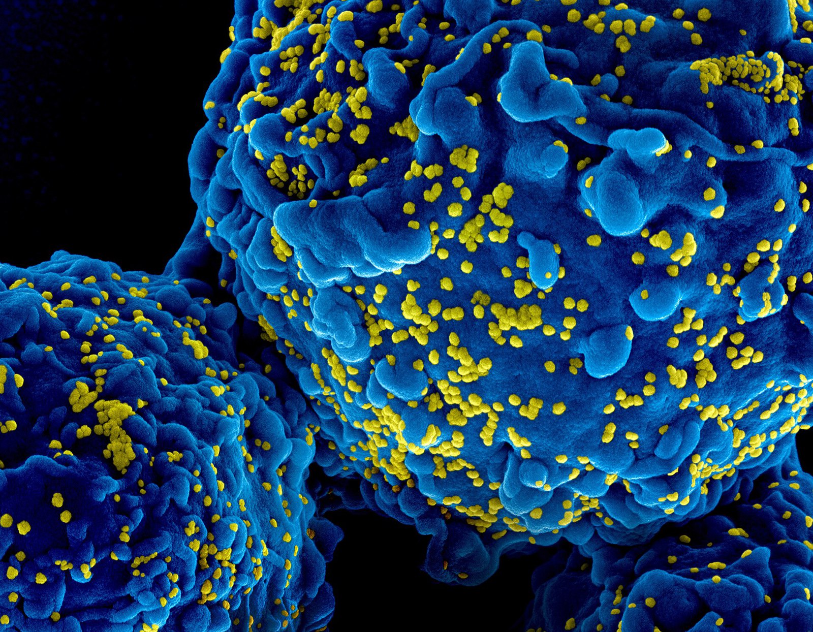
HIV-1 virus particles colorized scanning electron micrograph - 211
64 HIV-1 virus particles colorized scanning electron micrograph - 211
NIAID · CC BY 2.0 · sourceDownloadFull resolution (3406×2648) ↗
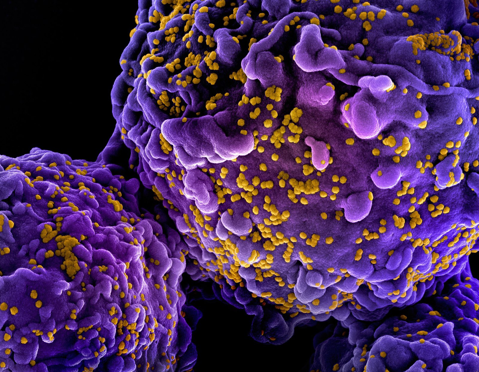
HIV-1 virus particles colorized scanning electron micrograph - 311
65 HIV-1 virus particles colorized scanning electron micrograph - 311
NIAID · CC BY 2.0 · sourceDownloadFull resolution (3406×2648) ↗
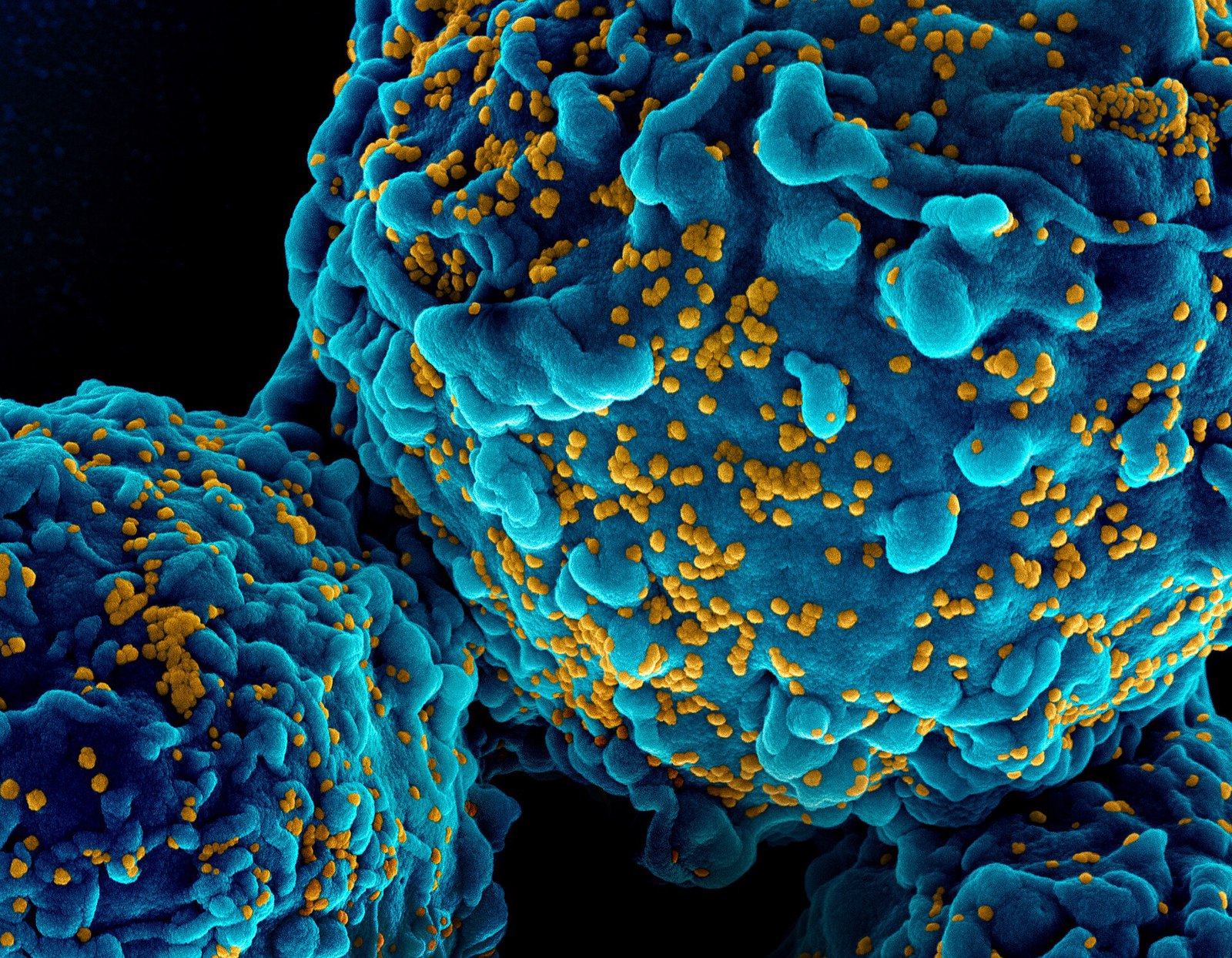
HIV-1 virus particles colorized scanning electron micrograph - 411
66 HIV-1 virus particles colorized scanning electron micrograph - 411
NIAID · CC BY 2.0 · sourceDownloadFull resolution (3406×2648) ↗
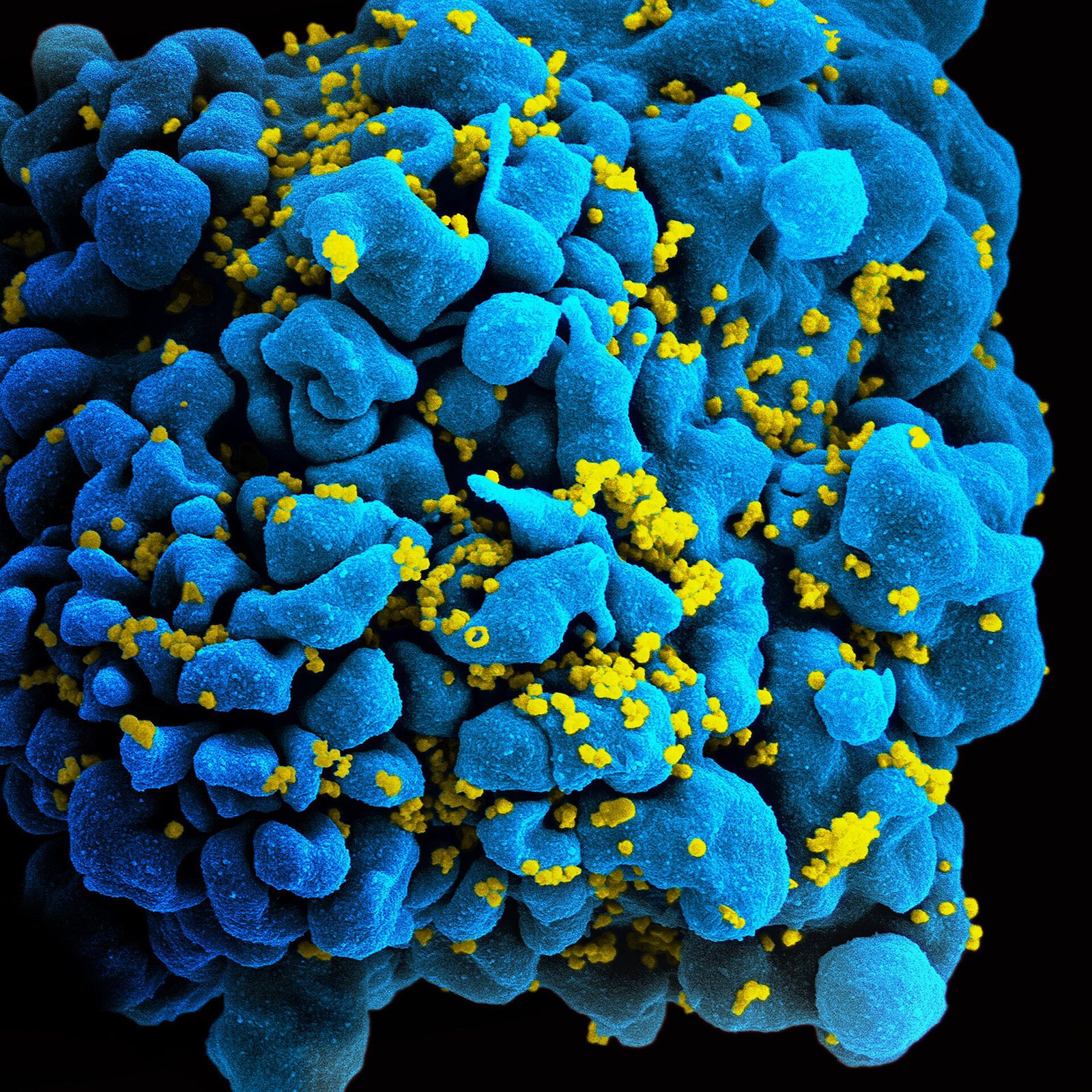
HIV-infected T cell (6813384933)
67 HIV-infected T cell (6813384933)
NIAID · CC BY 2.0 · sourceDownloadFull resolution (2560×2560) ↗
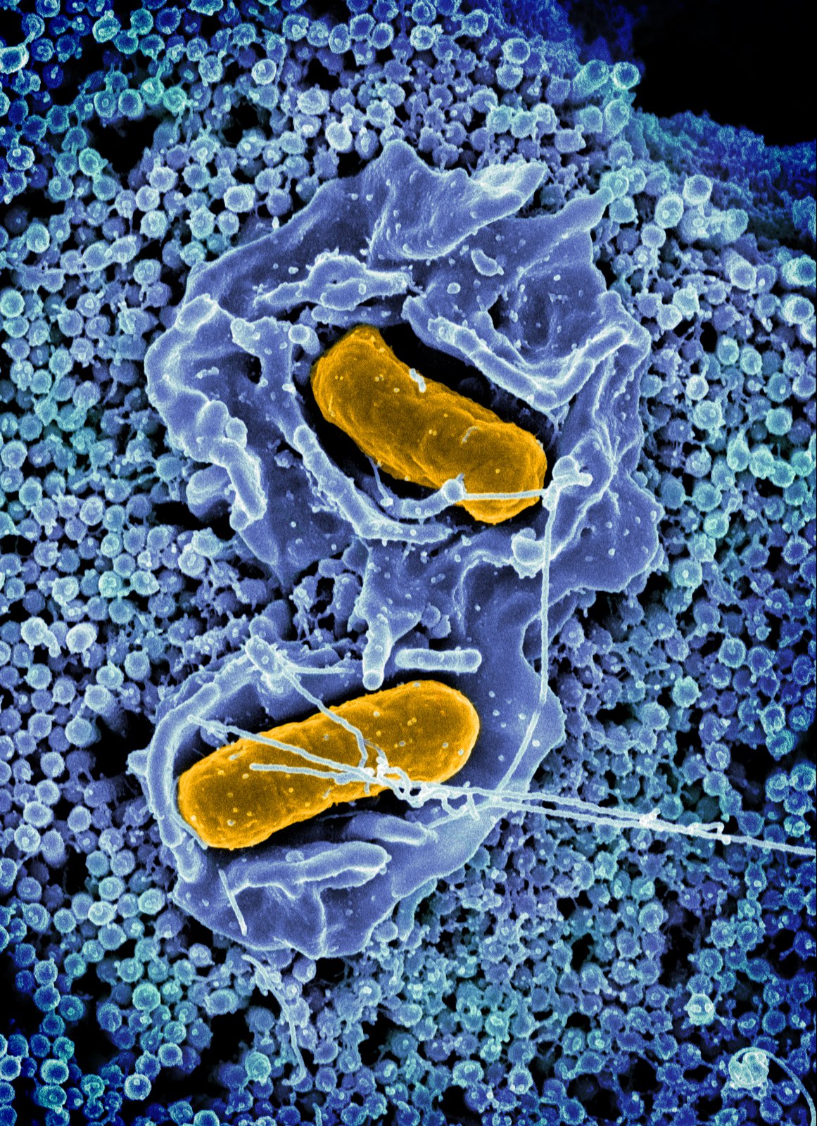
Salmonella Typhimurium invading a human epithelial cell - Scanning electron micrograph
68 Salmonella Typhimurium invading a human epithelial cell - Scanning electron micrograph
NIAID · CC BY 2.0 · sourceDownloadFull resolution (1764×2432) ↗
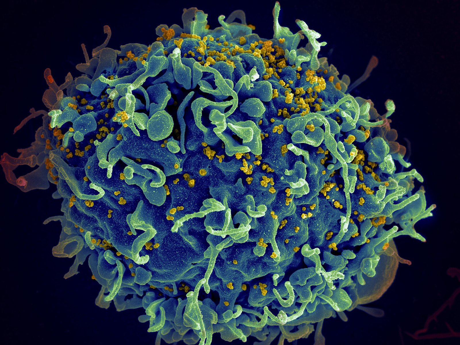
Scanning electron micrograph of a human H9 T cell infected with HIV virus particles
69 Scanning electron micrograph of a human H9 T cell infected with HIV virus particles
NIAID · CC BY 2.0 · sourceDownloadFull resolution (2361×1771) ↗
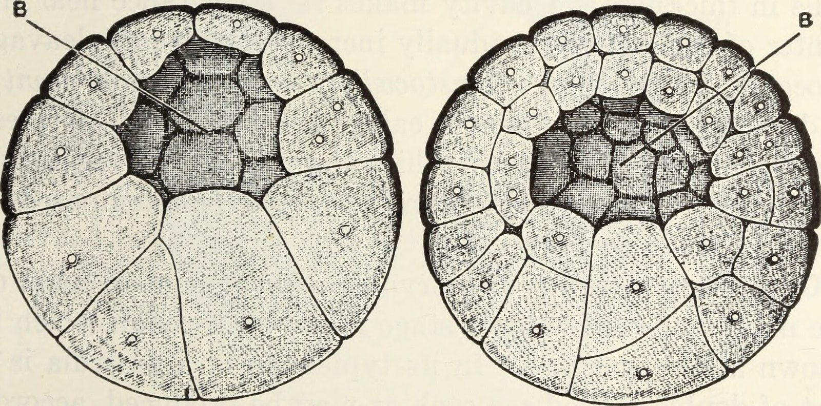
Frog blastula blastocoel
70 Frog blastula blastocoel
Holmes, Samuel J. · Public domain · sourceDownloadFull resolution (2208×1092) ↗
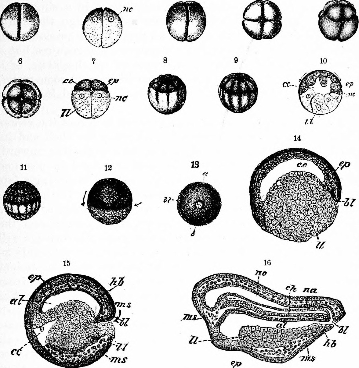
Frog embryo cleavage stadia
71 Frog embryo cleavage stadia
Mills, Wesley, 1847-1915 · Public domain · sourceDownloadFull resolution (1176×1208) ↗
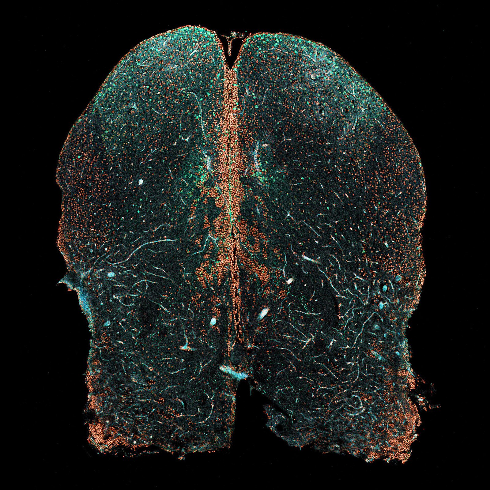
Corte cerebro pez cebra visto con microscopio confocal
72 Corte cerebro pez cebra visto con microscopio confocal
Brandon Antonio Segura Torres & Priscilla Vieto Bonilla · CC BY-SA 4.0 · sourceDownloadFull resolution (4069×4069) ↗
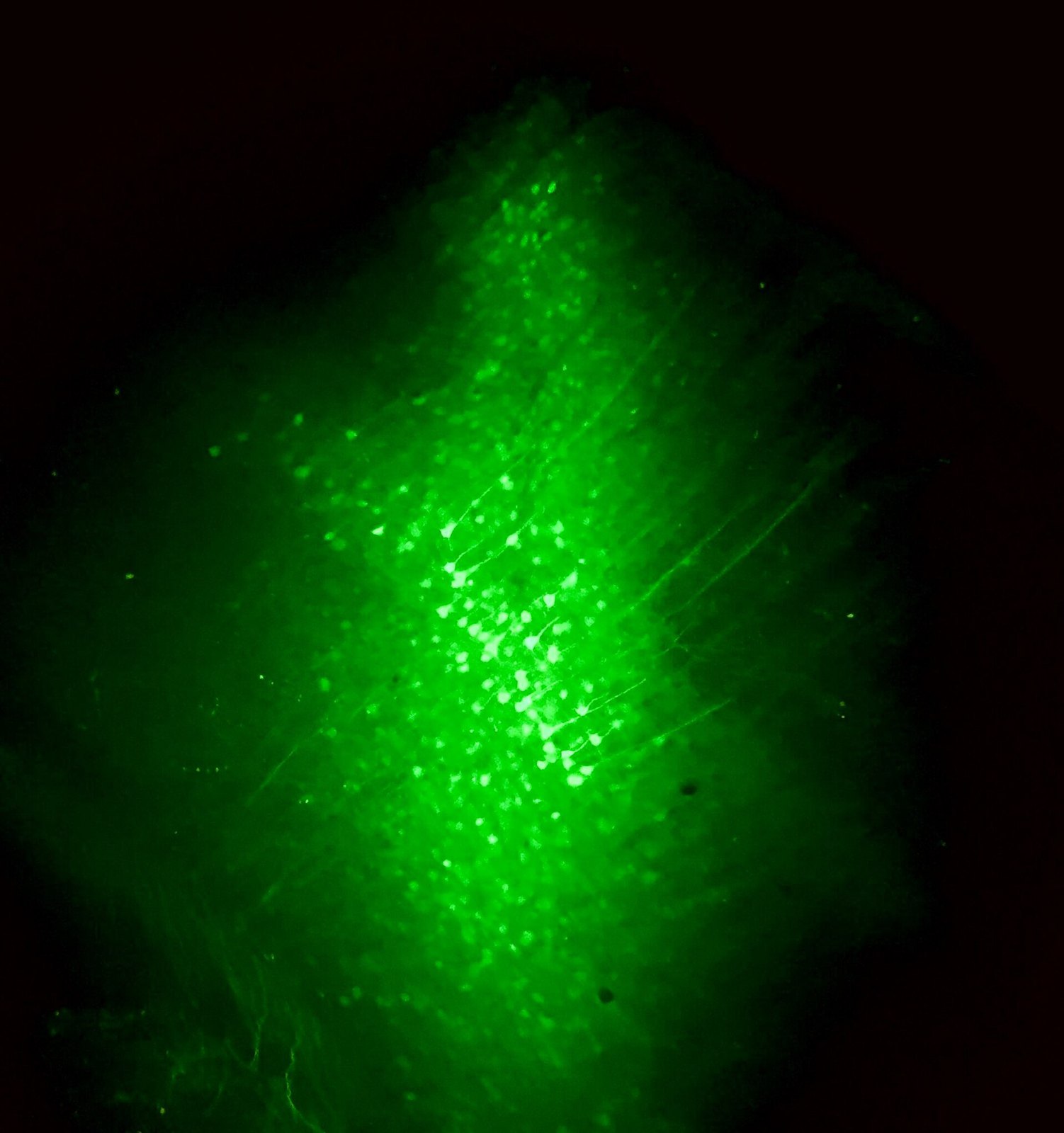
Cortical neurons
73 Cortical neurons
Luisa Demarchi, Ilaria Bertocchi, Alessandra Oberto · CC BY-SA 4.0 · sourceDownloadFull resolution (2253×2400) ↗
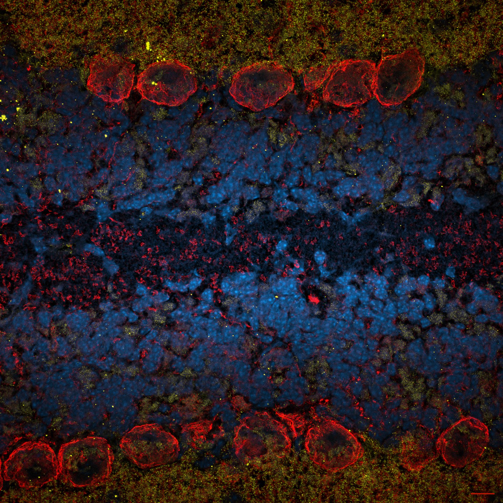
Purkinje neuron cell bodies in a mouse cerebellum fold - Ksena Longrin
74 Purkinje neuron cell bodies in a mouse cerebellum fold - Ksena Longrin
Morne Arin · CC BY 4.0 · sourceDownloadFull resolution (3082×3082) ↗
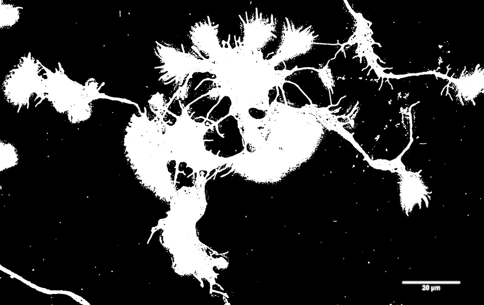
SUM 110913 Cort Neurons 2.5d in vitro 488 Phalloidin no perm 4 cmle-2
75 SUM 110913 Cort Neurons 2.5d in vitro 488 Phalloidin no perm 4 cmle-2
Howard Vindin · CC BY-SA 4.0 · sourceDownloadFull resolution (4096×2584) ↗
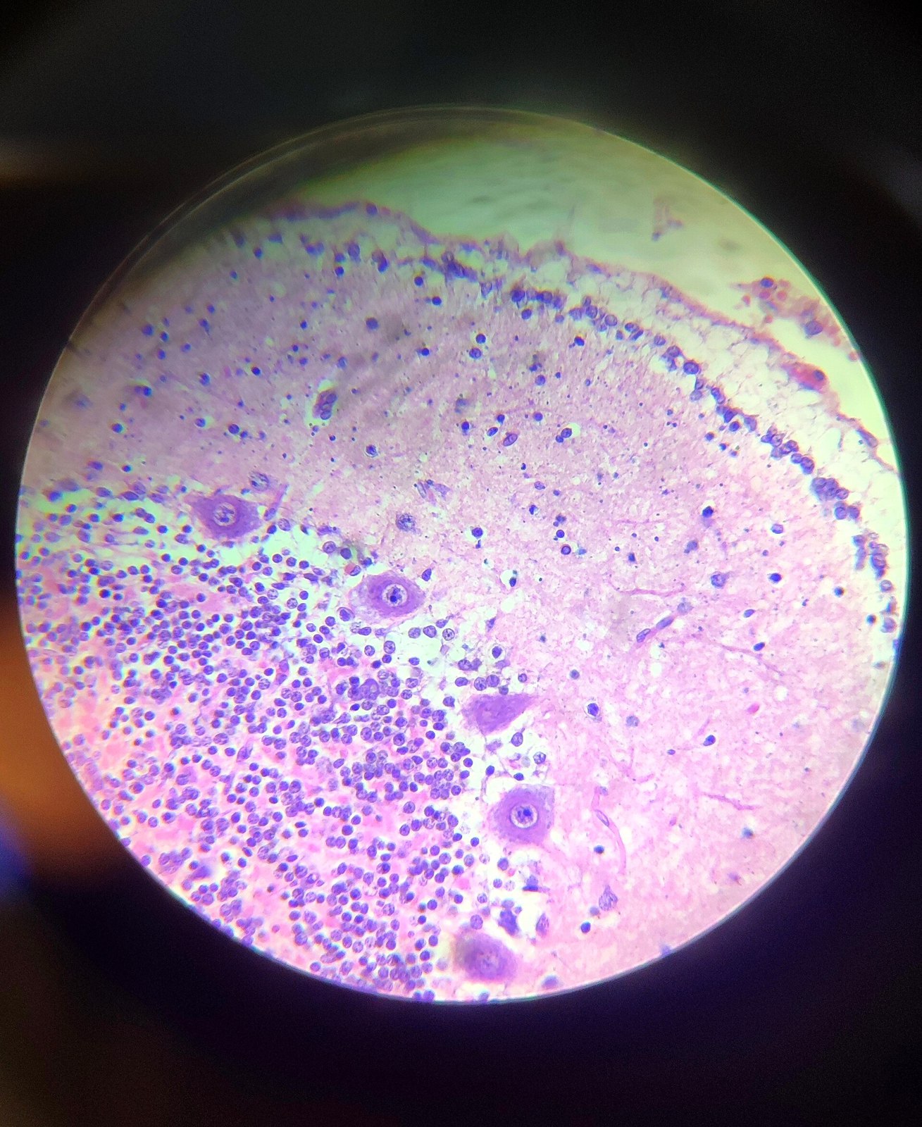
Mouse cerebellar cortex H&E 40x
76 Mouse cerebellar cortex H&E 40x
Hesperides29 · CC BY-SA 4.0 · sourceDownloadFull resolution (2426×2966) ↗
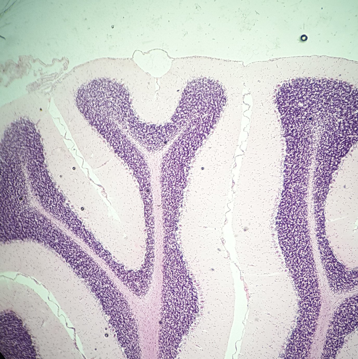
Мозжечковая кора кошки. Окраска гематоксилин-эозин. Увеличение x100
77 Мозжечковая кора кошки. Окраска гематоксилин-эозин. Увеличение x100
Polina DTsareva · CC BY 4.0 · sourceDownloadFull resolution (1377×1378) ↗
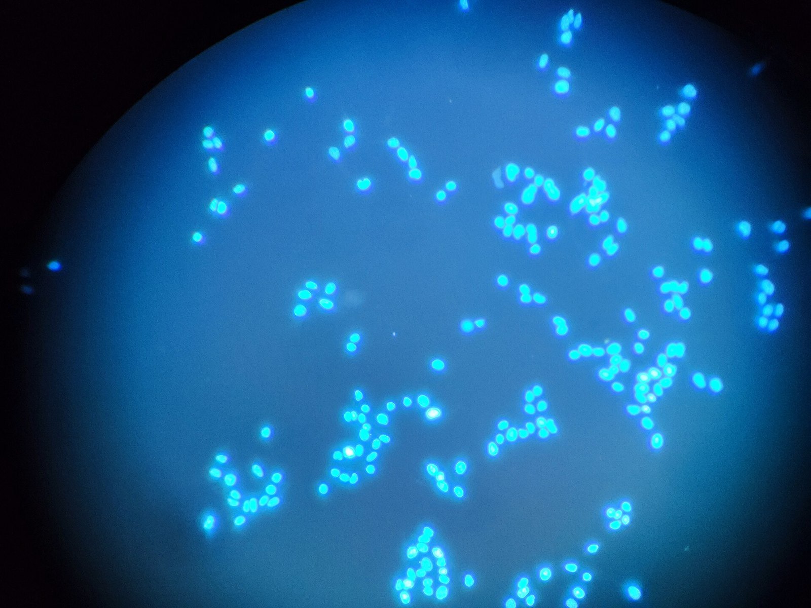
Fluorescence microscopy of the DNA of the oral cancer cells
78 Fluorescence microscopy of the DNA of the oral cancer cells
Korinna · CC BY 4.0 · sourceDownloadFull resolution (4096×3072) ↗
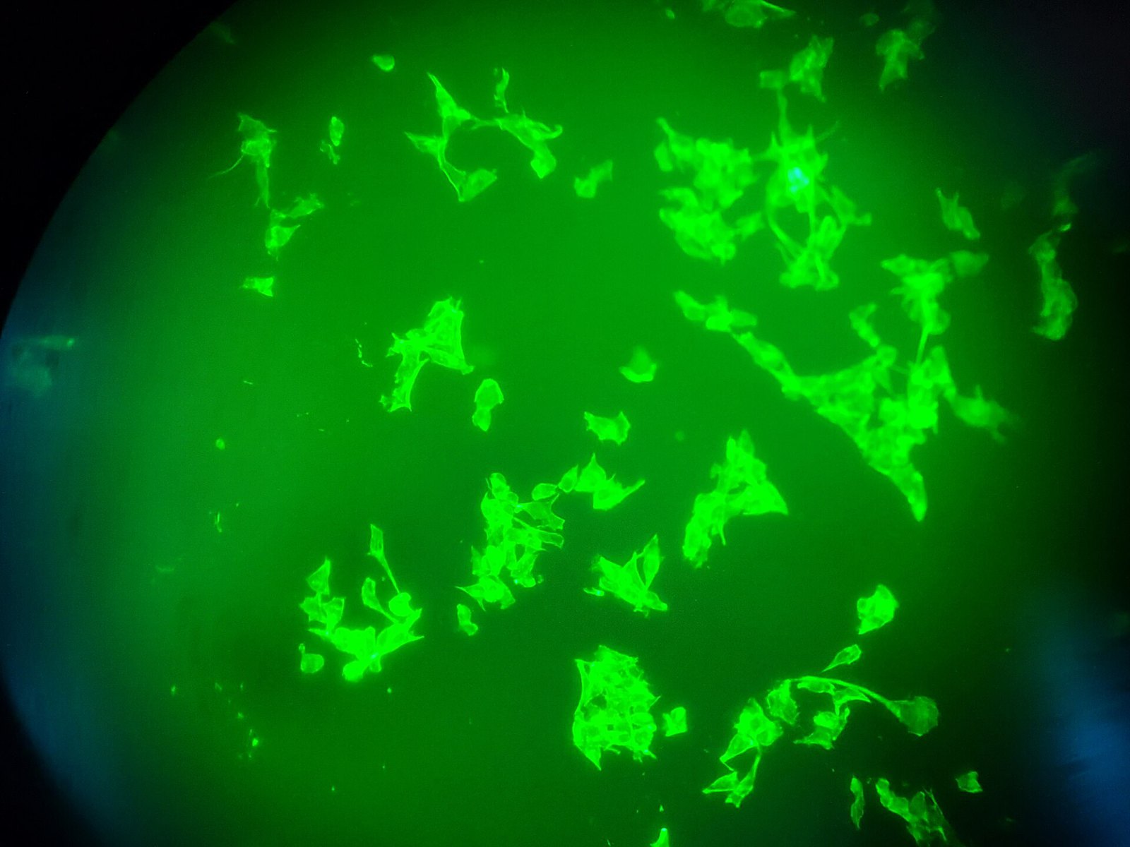
Fluorescence microscopy of the oral cancer cells
79 Fluorescence microscopy of the oral cancer cells
Korinna · CC BY 4.0 · sourceDownloadFull resolution (4096×3072) ↗
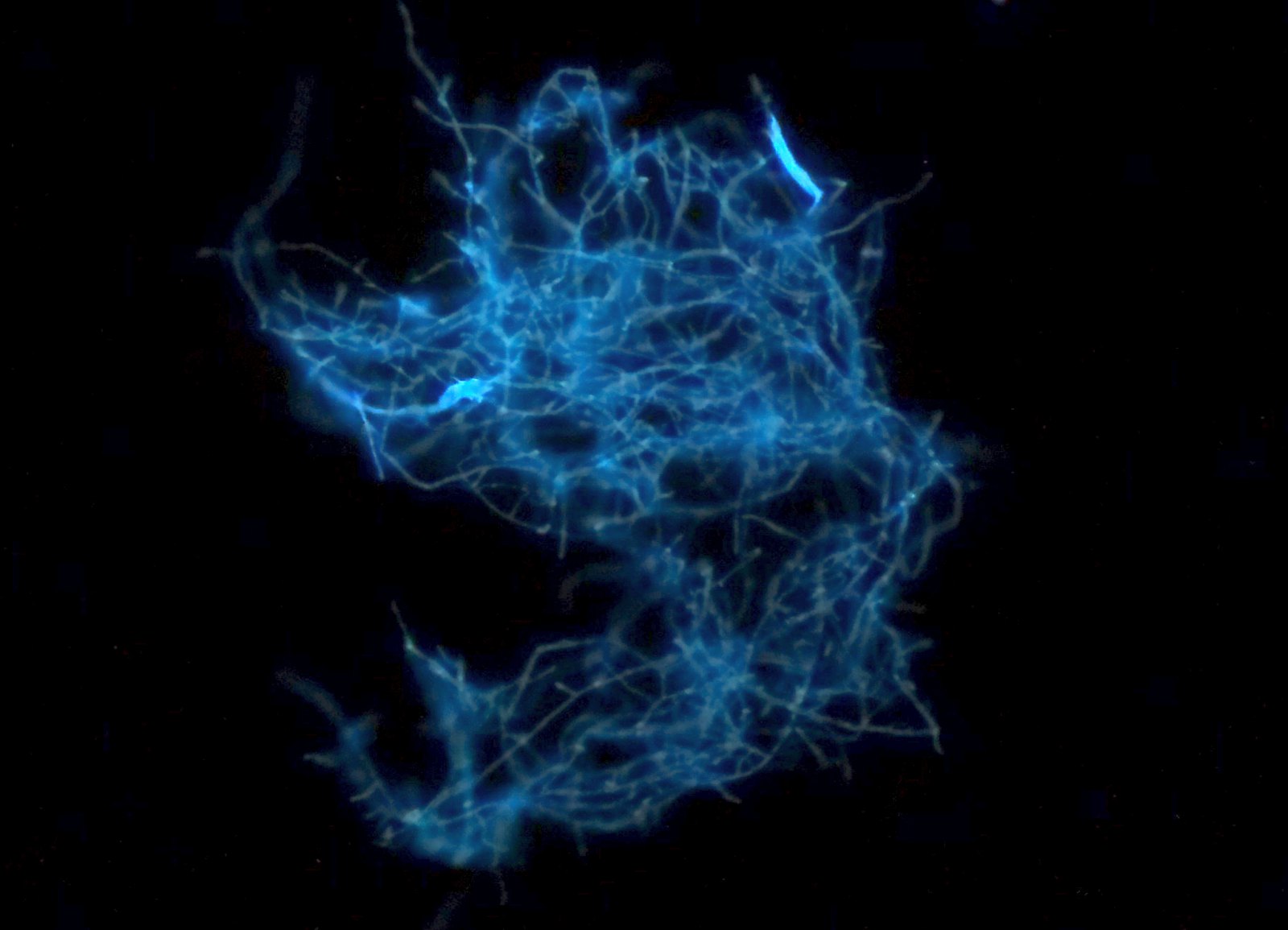
Fungi seen with fluorescence microscope
80 Fungi seen with fluorescence microscope
Juan Carlos Fonseca Mata · CC BY 4.0 · sourceDownloadFull resolution (1814×1310) ↗
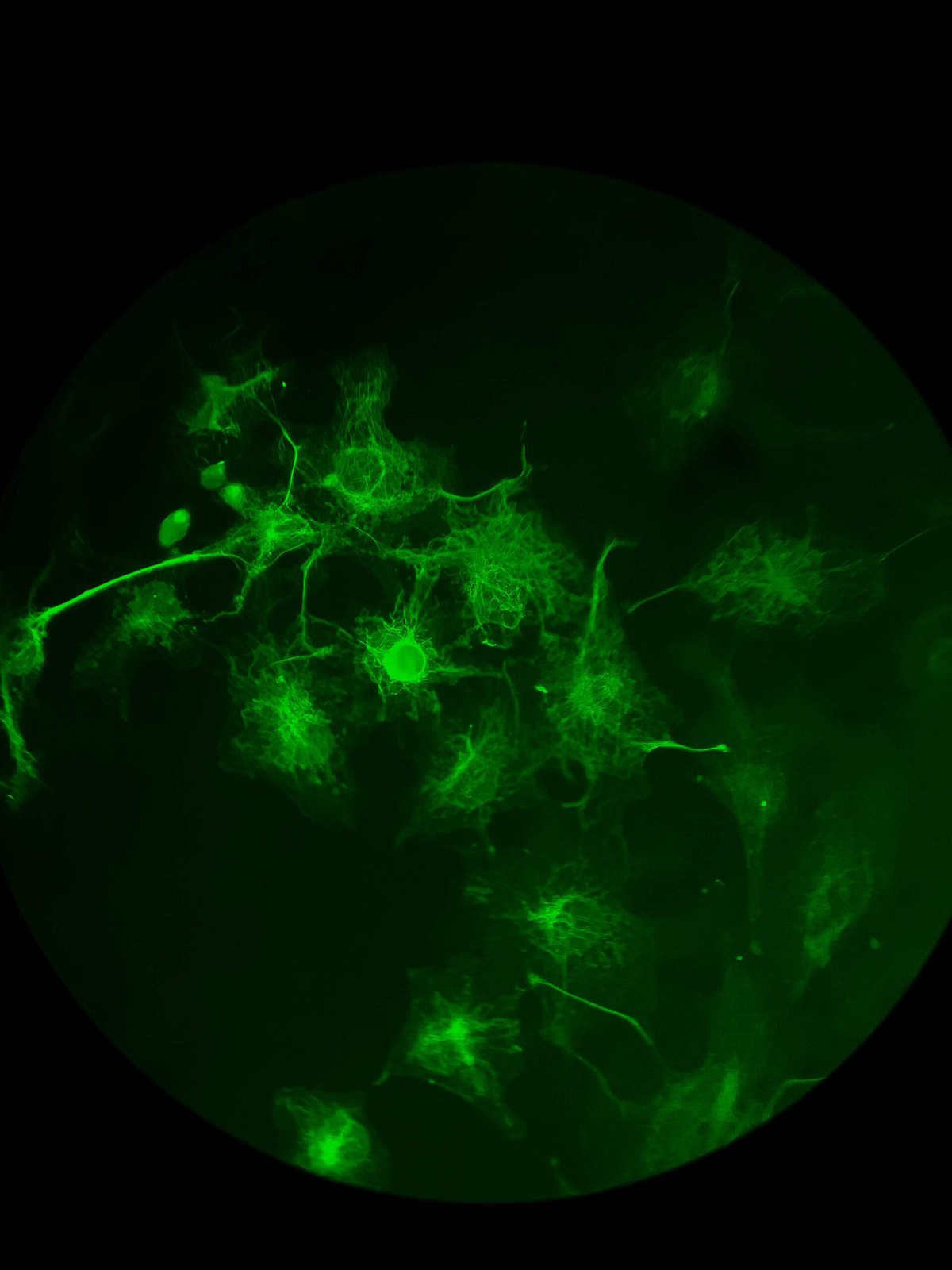
Green Fluorescence Microscopy
81 Green Fluorescence Microscopy
Jamil Baza · CC BY 4.0 · sourceDownloadFull resolution (3024×4032) ↗
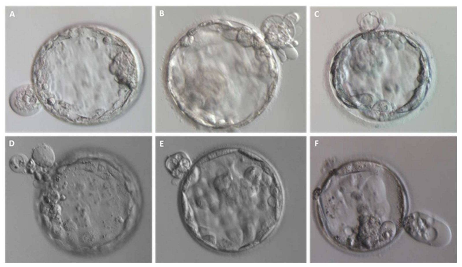
Entire blastomeres are excluded upon blastocyst formation
82 Entire blastomeres are excluded upon blastocyst formation
Cecchele, A.; Cermisoni, G.C.; Giacomini, E.; Pinna, M.; Vigano, P. · CC BY 4.0 · sourceDownloadFull resolution (3130×1809) ↗
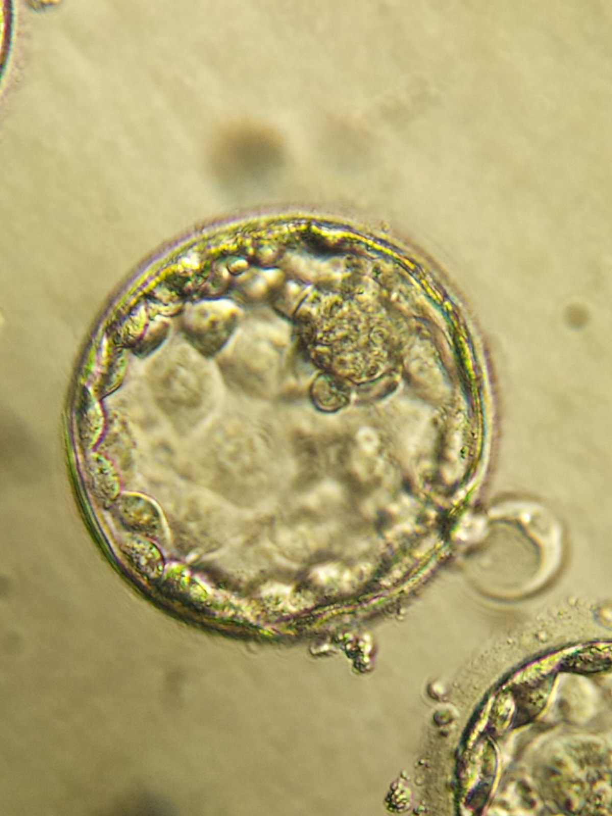
Inbound2890638149762117997
83 Inbound2890638149762117997
Kurtyak · CC BY 4.0 · sourceDownloadFull resolution (3000×4000) ↗
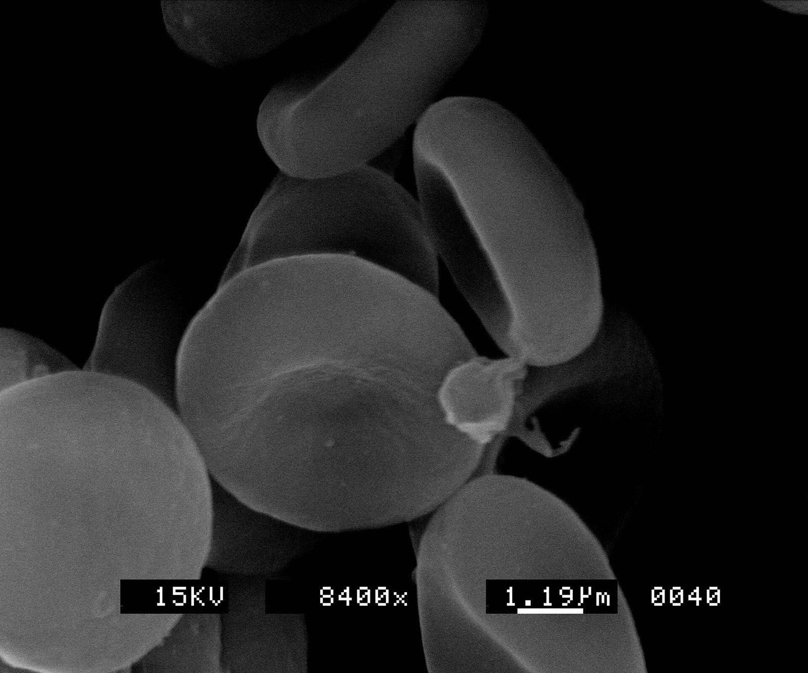
Red blood cells (2)
84 Red blood cells (2)
Scootdive (talk · contribs) · CC BY-SA 3.0 · sourceDownloadFull resolution (2400×2000) ↗
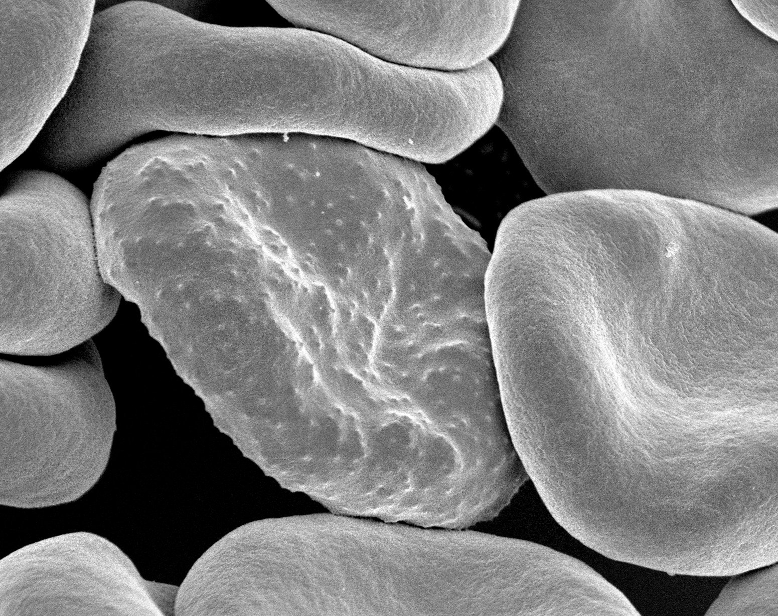
Red blood cells infected with malaria
85 Red blood cells infected with malaria
Rick Fairhurst and Jordan Zuspann, National Institute of Allergy and Infectious Diseases, National Institutes of Health · Public domain · sourceDownloadFull resolution (1551×1229) ↗
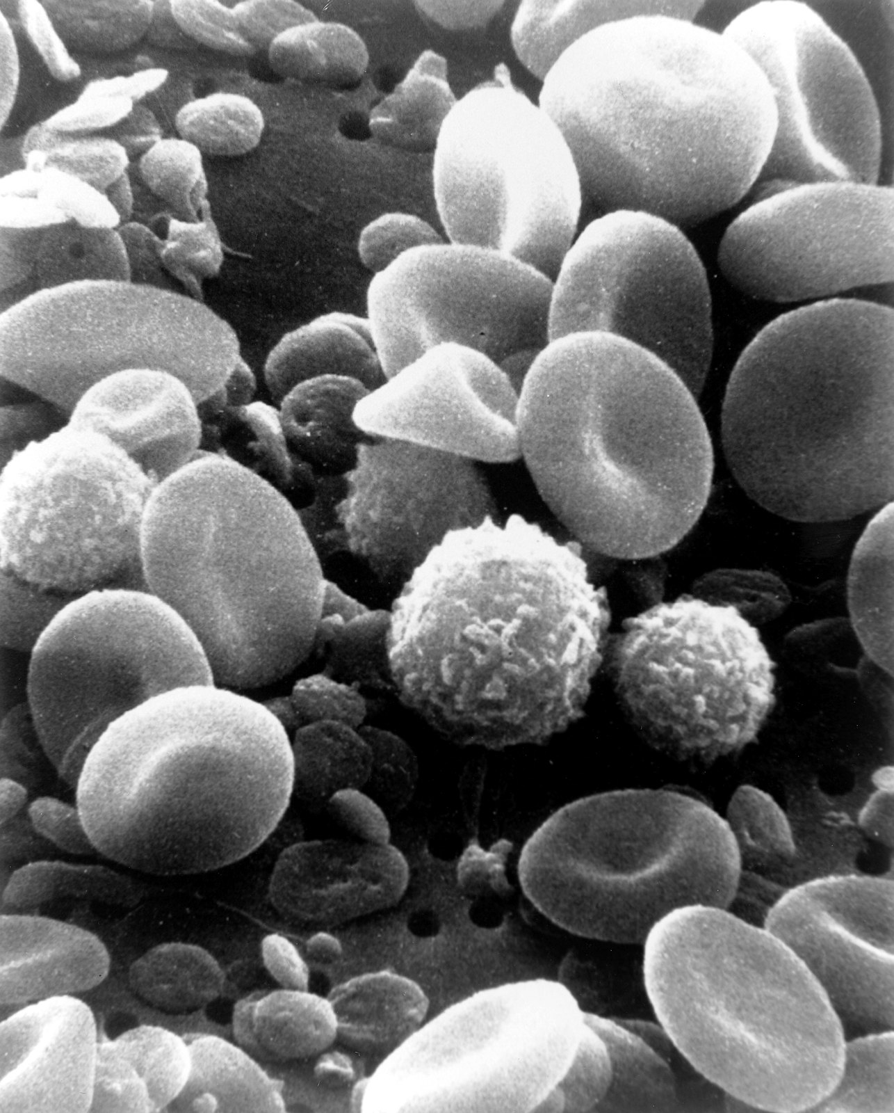
SEM blood cells
86 SEM blood cells
Bruce Wetzel (photographer). Harry Schaefer (photographer) · Public domain · sourceDownloadFull resolution (1800×2239) ↗
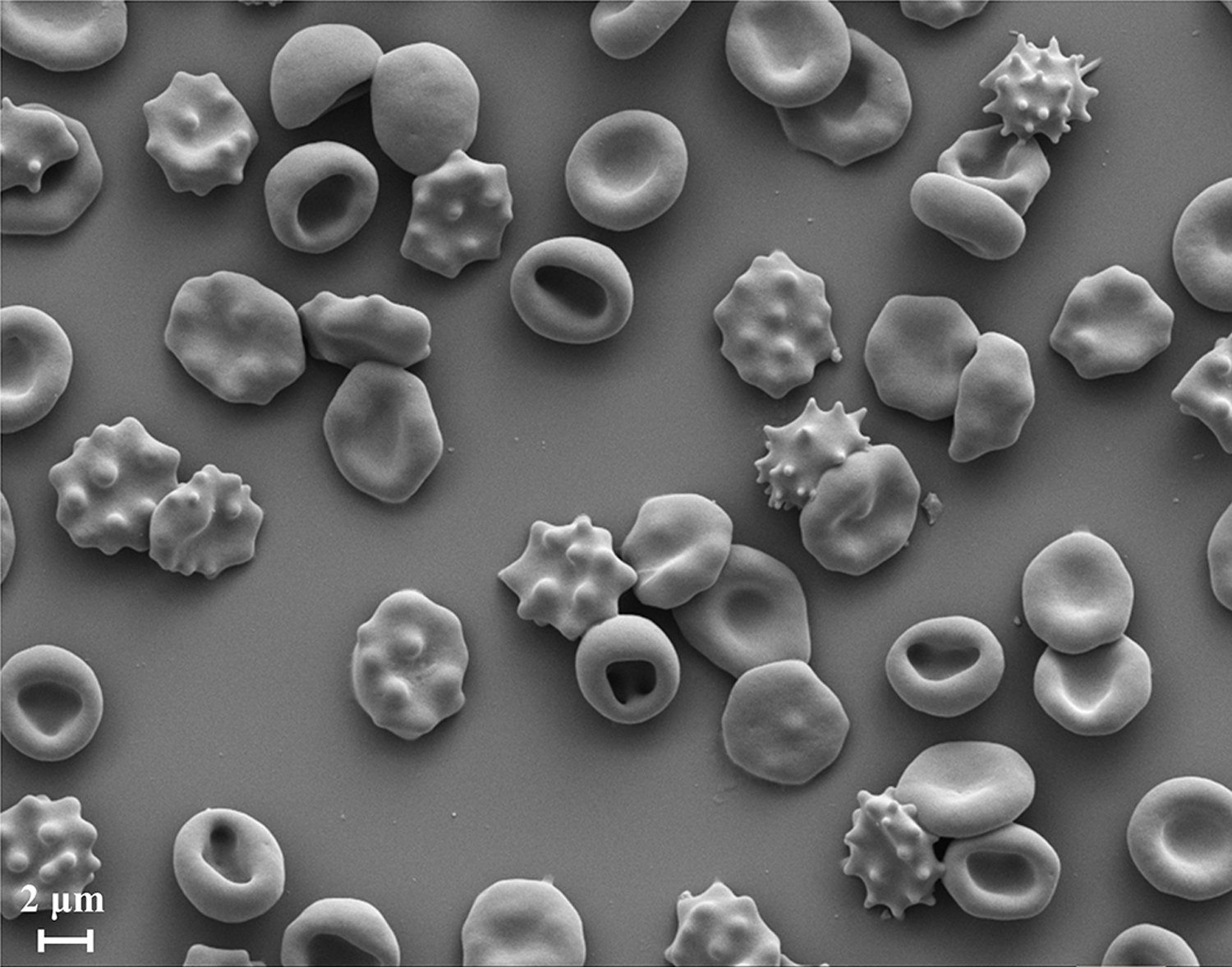
Scanning electron microscope (SEM) image of a RBC population having different morphology stages of stomatocytosis and echinocytosis
87 Scanning electron microscope (SEM) image of a RBC population having different morphology stages of stomatocytosis and echinocytosis
Geekiyanage NM, Balanant MA, Sauret E, Saha S, Flower R, Lim CT, et al. · CC BY-SA 4.0 · sourceDownloadFull resolution (1500×1177) ↗
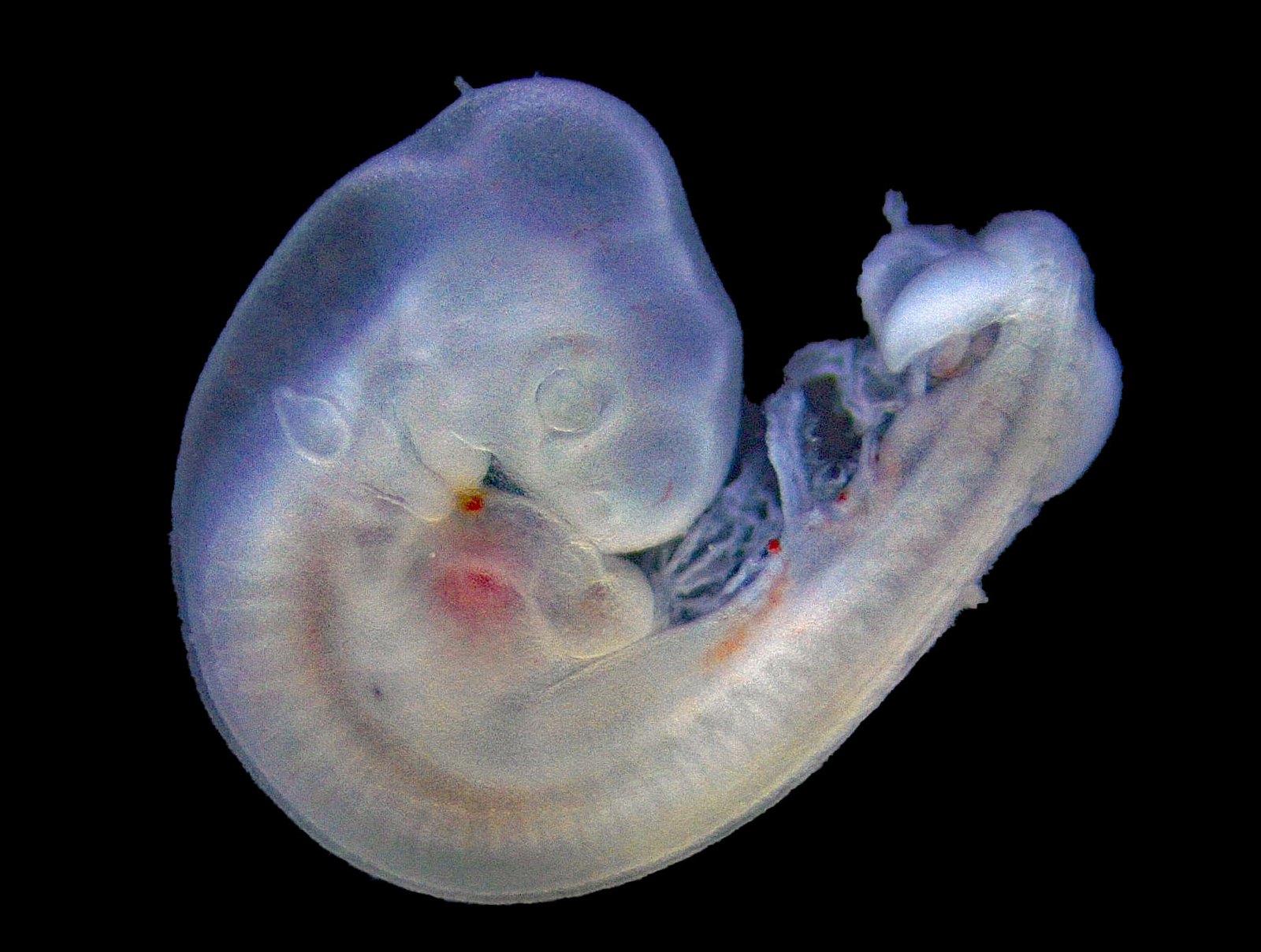
Chick embryo 3d
88 Chick embryo 3d
PaulT (Gunther Tschuch) · CC BY-SA 4.0 · sourceDownloadFull resolution (1780×1345) ↗
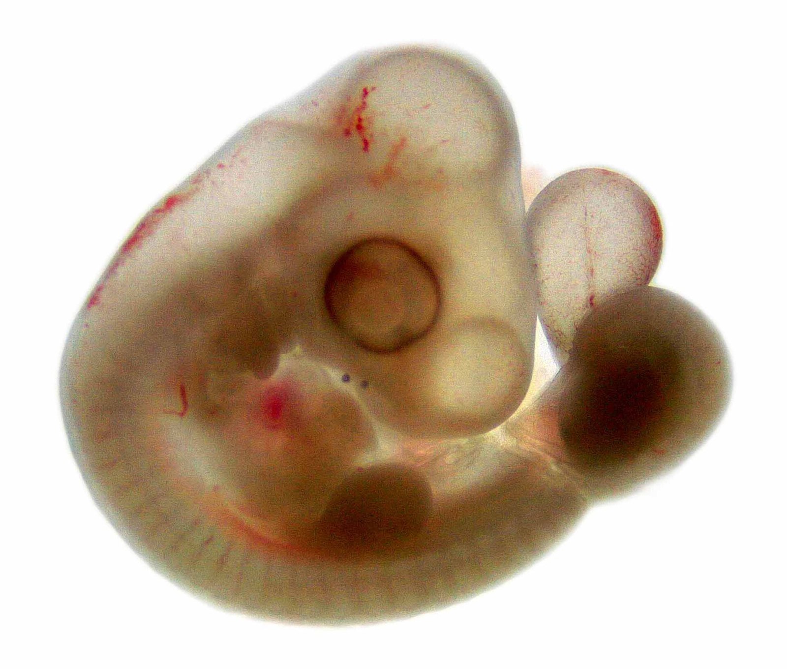
Chick embryo 4d
89 Chick embryo 4d
PaulT (Gunther Tschuch) · CC BY-SA 4.0 · sourceDownloadFull resolution (1926×1637) ↗
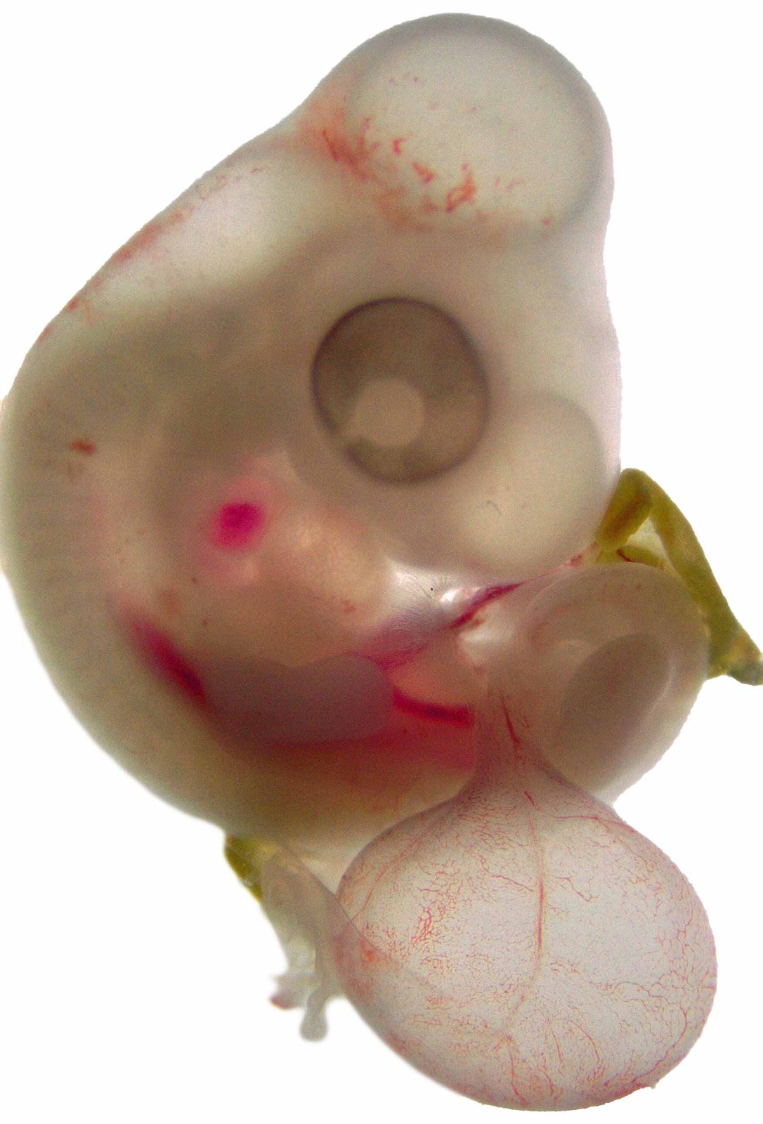
Chick embryo 5d
90 Chick embryo 5d
PaulT (Gunther Tschuch) · CC BY-SA 4.0 · sourceDownloadFull resolution (1749×2576) ↗
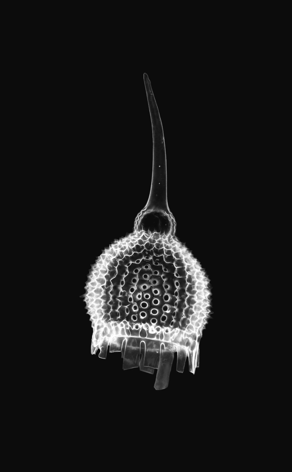
Anthocyrtium hispidum Haeckel - Radiolarian (34986365113)
91 Anthocyrtium hispidum Haeckel - Radiolarian (34986365113)
Picturepest · CC BY 2.0 · sourceDownloadFull resolution (4252×6863) ↗
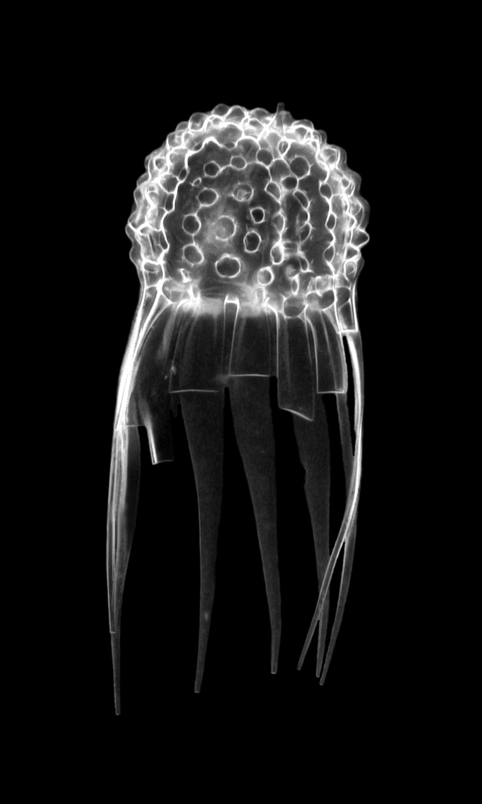
Caclocyma ( = Halicalyptra) petalospyris HAECKEL - Radiolarian (31708336584)
92 Caclocyma ( = Halicalyptra) petalospyris HAECKEL - Radiolarian (31708336584)
Picturepest · CC BY 2.0 · sourceDownloadFull resolution (1200×2000) ↗
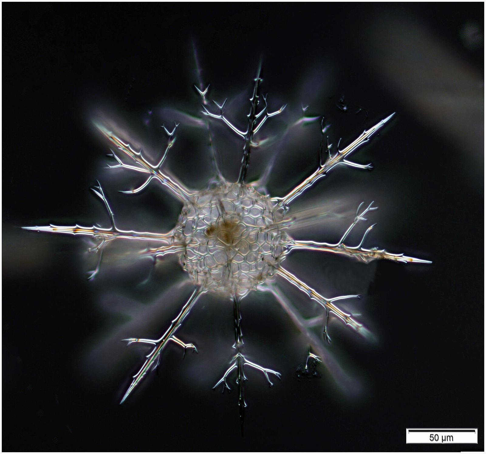
Cladococcus abietinus
93 Cladococcus abietinus
tintinnidguy · CC BY 2.0 · sourceDownloadFull resolution (2724×2548) ↗
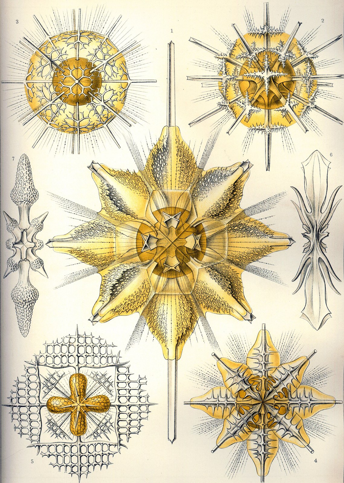
Haeckel Acanthometra
94 Haeckel Acanthometra
Ernst Haeckel · Public domain · sourceDownloadFull resolution (2346×3294) ↗
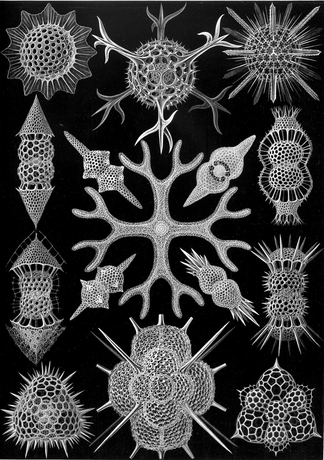
Haeckel Spumellaria
95 Haeckel Spumellaria
Ernst Haeckel · Public domain · sourceDownloadFull resolution (2344×3324) ↗
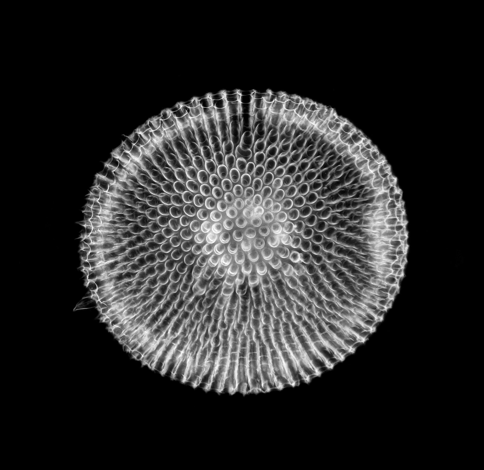
Phacodiscus clypeus Haeckel - Radiolarian (36086846420)
96 Phacodiscus clypeus Haeckel - Radiolarian (36086846420)
Picturepest · CC BY 2.0 · sourceDownloadFull resolution (3500×3400) ↗
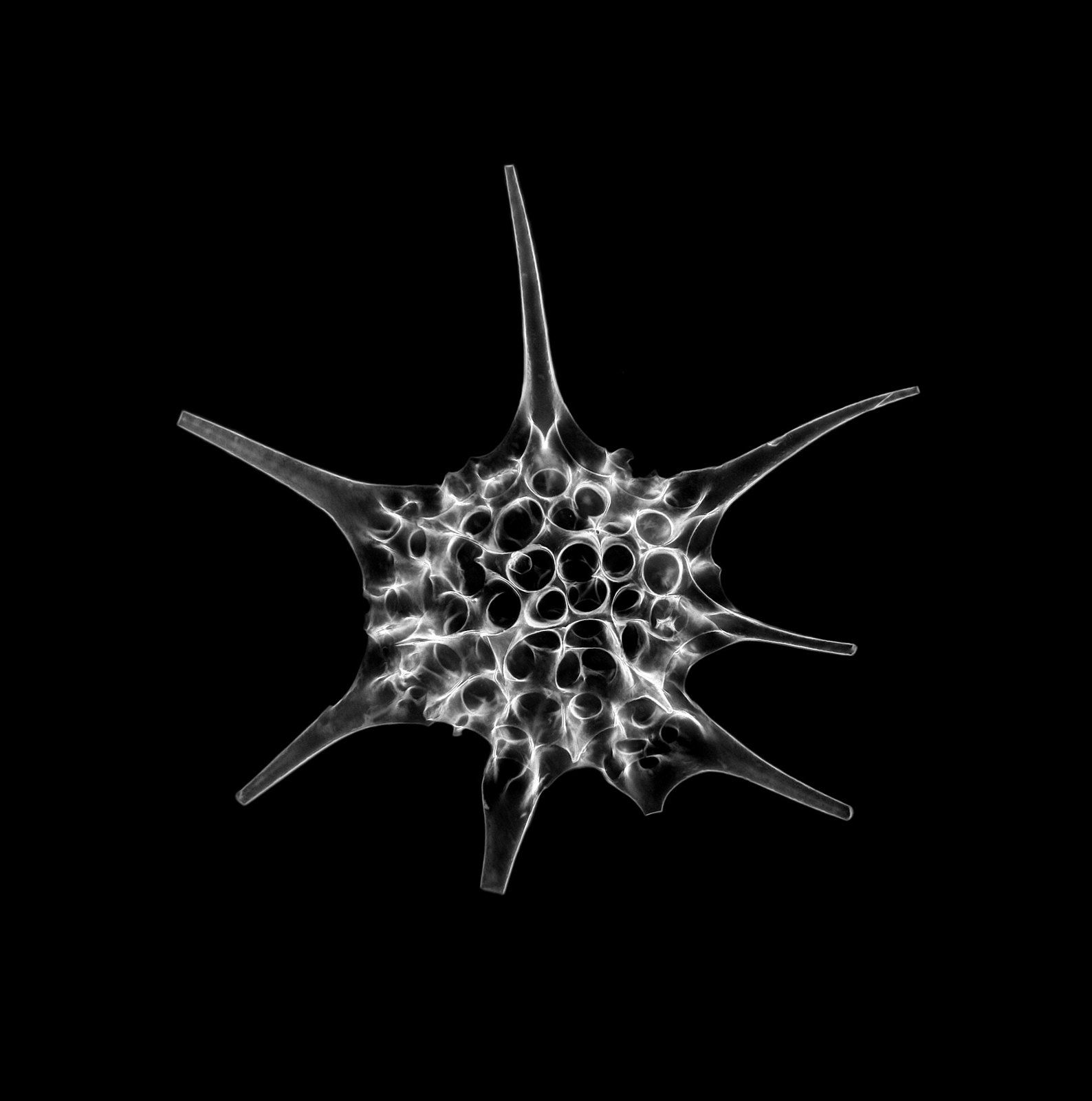
Radiolarian - Dorataspida sp
97 Radiolarian - Dorataspida sp
Andreas Drews · CC BY 2.0 · sourceDownloadFull resolution (5160×5200) ↗
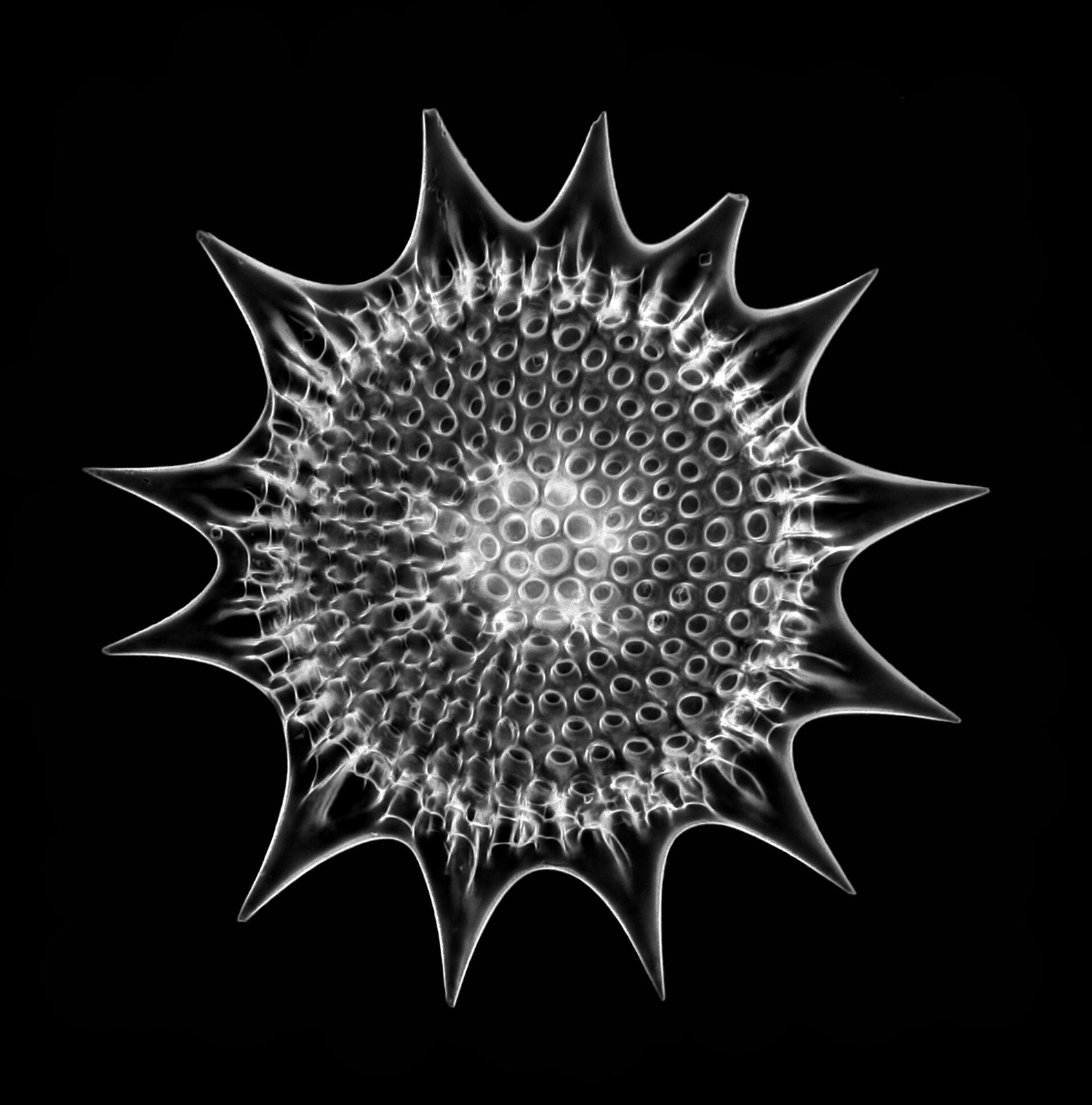
Radiolarian - Heliodiscus umbonatus (Ehr.), Haeckel (28187768550)
98 Radiolarian - Heliodiscus umbonatus (Ehr.), Haeckel (28187768550)
Picturepest · CC BY 2.0 · sourceDownloadFull resolution (2000×2024) ↗

Embryos & the womb
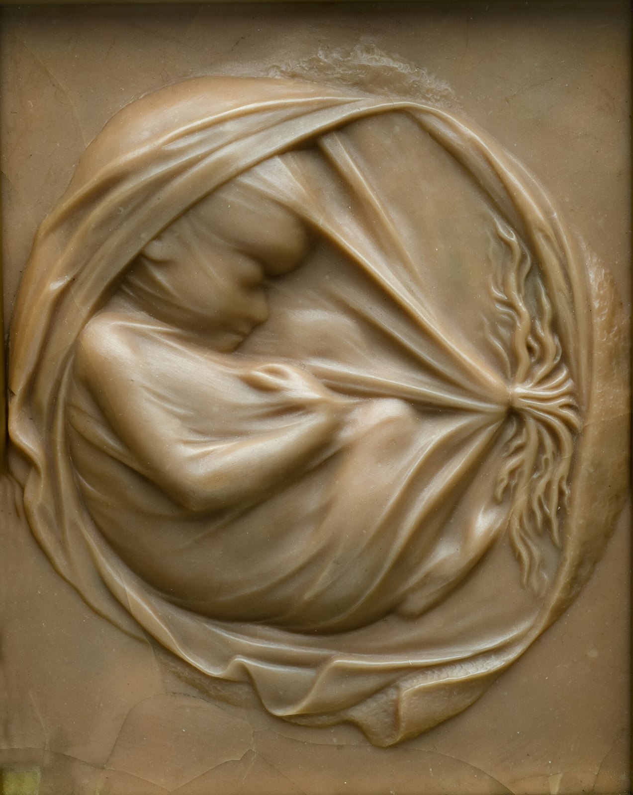
Foetus in placenta in utero. Wellcome L0057759
99 Foetus in placenta in utero. Wellcome L0057759
Unknown · CC BY 4.0 · sourceDownloadFull resolution (2953×3713) ↗
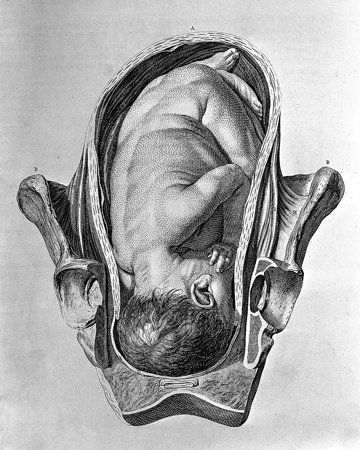
Foetus in utero. Wellcome L0004300
100 Foetus in utero. Wellcome L0004300
Unknown · CC BY 4.0 · sourceDownloadFull resolution (1215×1517) ↗
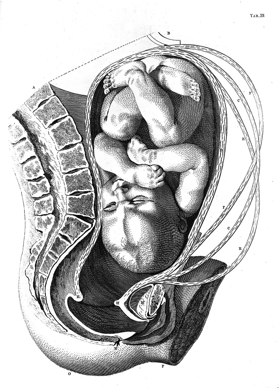
Foetus in utero. Wellcome L0013508
101 Foetus in utero. Wellcome L0013508
Unknown · CC BY 4.0 · sourceDownloadFull resolution (1154×1600) ↗
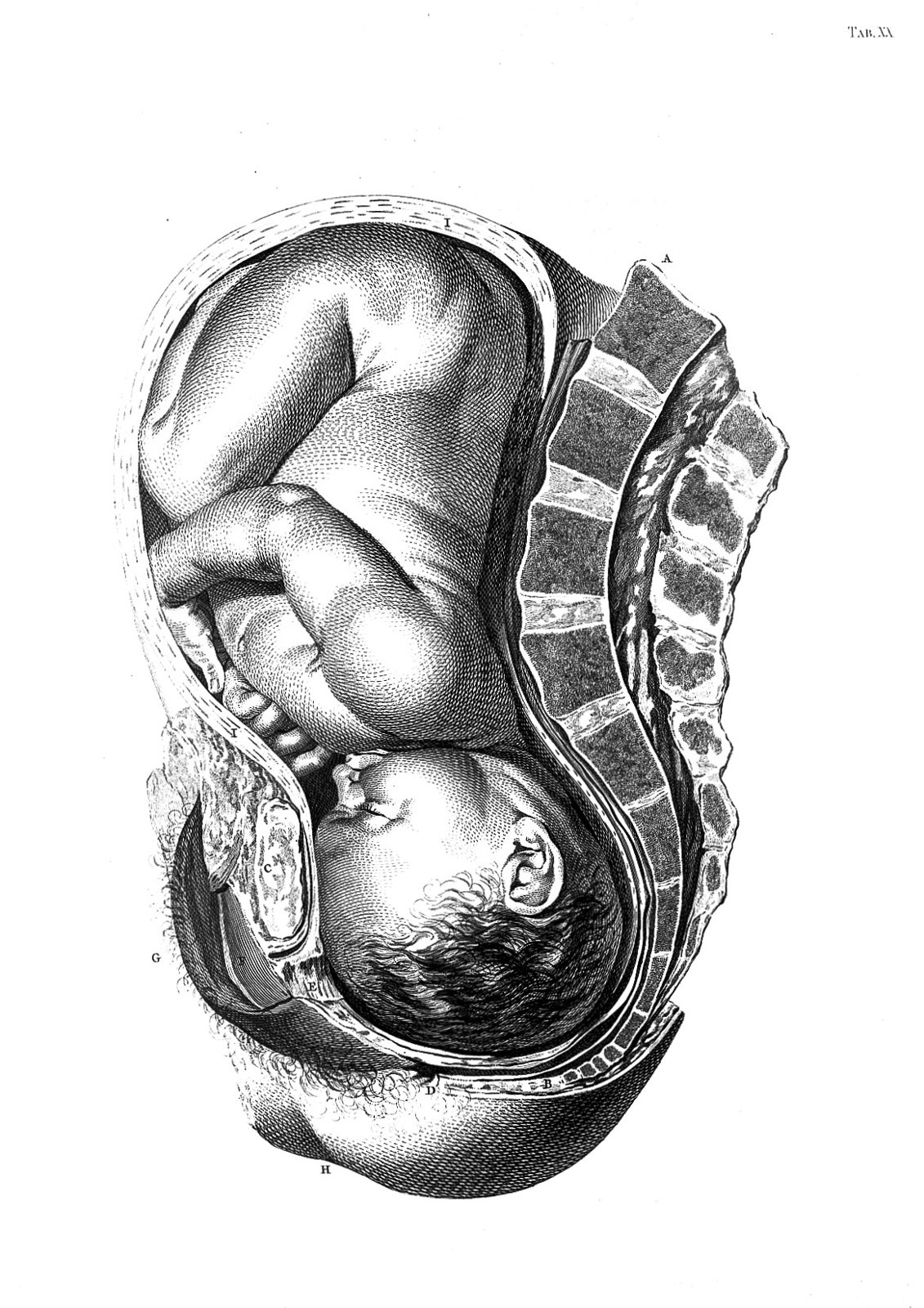
Foetus in utero. Wellcome L0013509
102 Foetus in utero. Wellcome L0013509
Unknown · CC BY 4.0 · sourceDownloadFull resolution (1166×1650) ↗
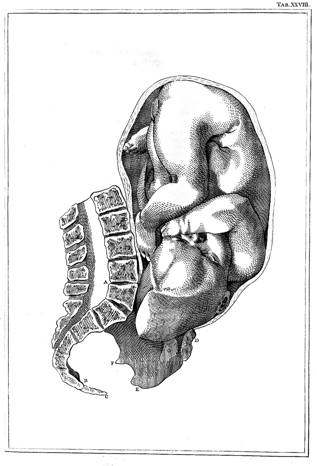
Foetus in utero. Wellcome L0013510
103 Foetus in utero. Wellcome L0013510
Unknown · CC BY 4.0 · sourceDownloadFull resolution (1113×1638) ↗
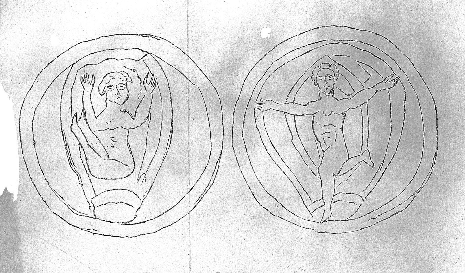
Foetus in utero. Wellcome L0016391
104 Foetus in utero. Wellcome L0016391
Unknown · CC BY 4.0 · sourceDownloadFull resolution (1814×1064) ↗
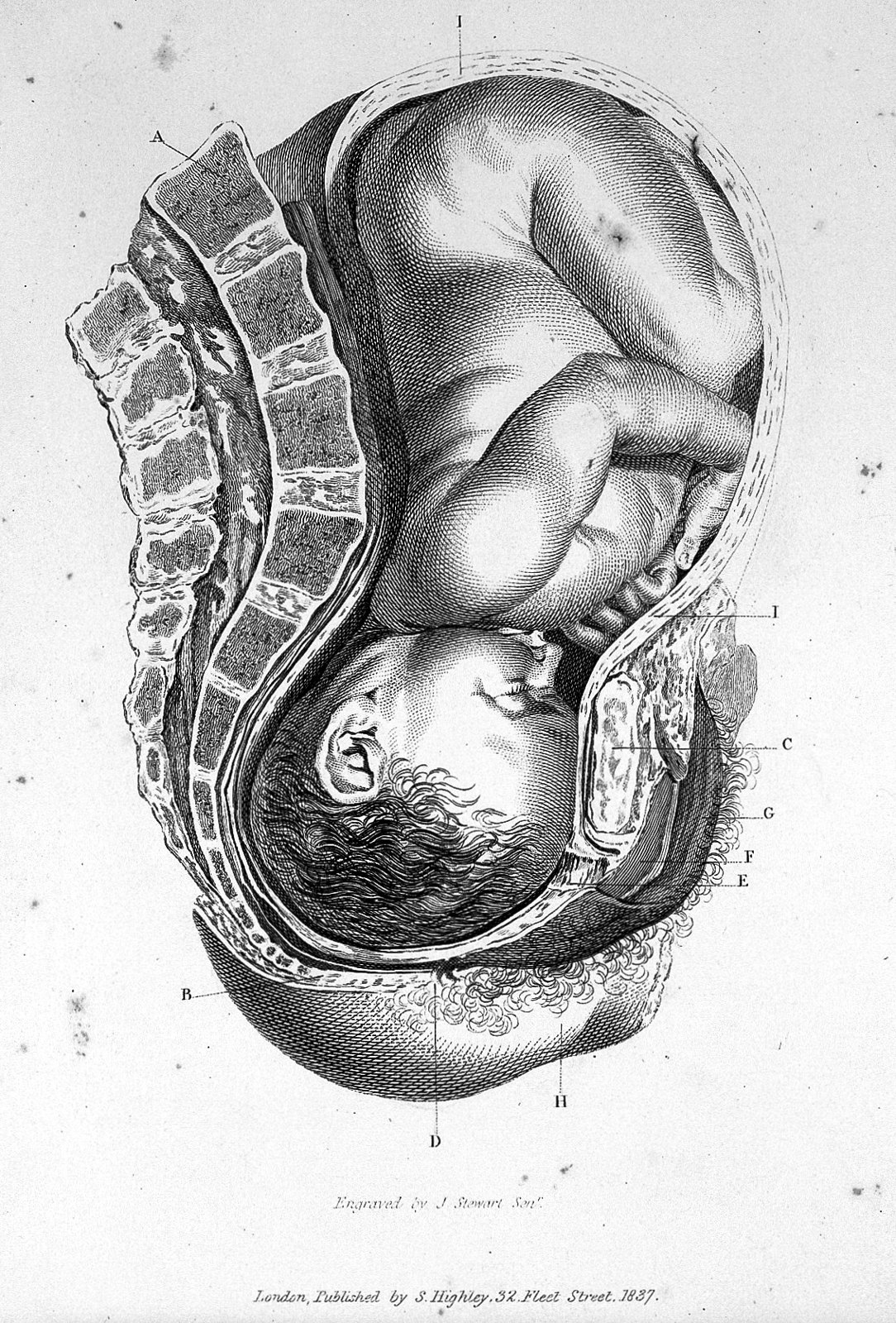
Foetus in utero. Wellcome L0018065
105 Foetus in utero. Wellcome L0018065
Unknown · CC BY 4.0 · sourceDownloadFull resolution (1134×1674) ↗
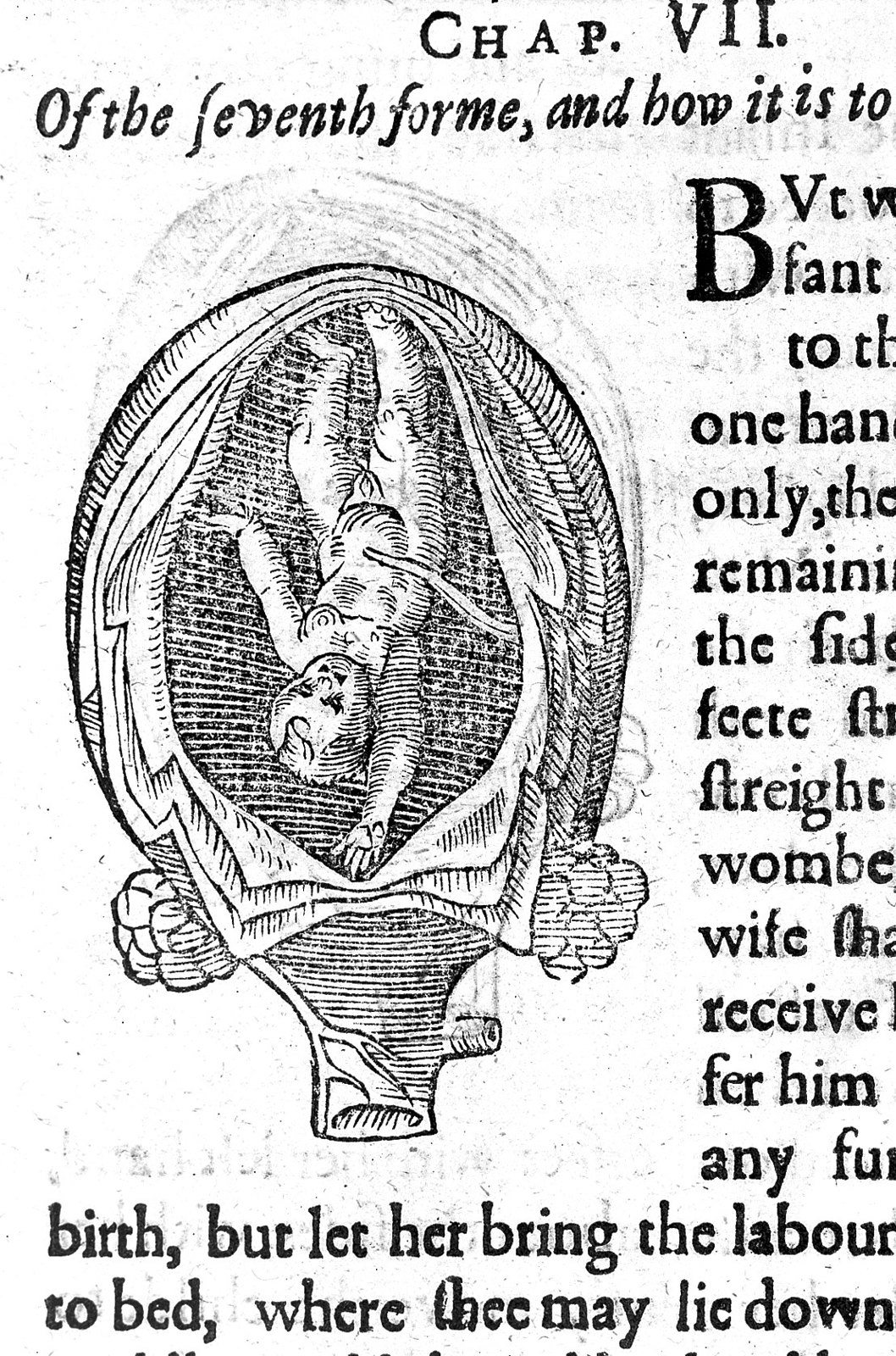
Foetus in utero from Rueff "Expert midwife" Wellcome L0014439
106 Foetus in utero from Rueff "Expert midwife" Wellcome L0014439
Unknown · CC BY 4.0 · sourceDownloadFull resolution (1108×1675) ↗
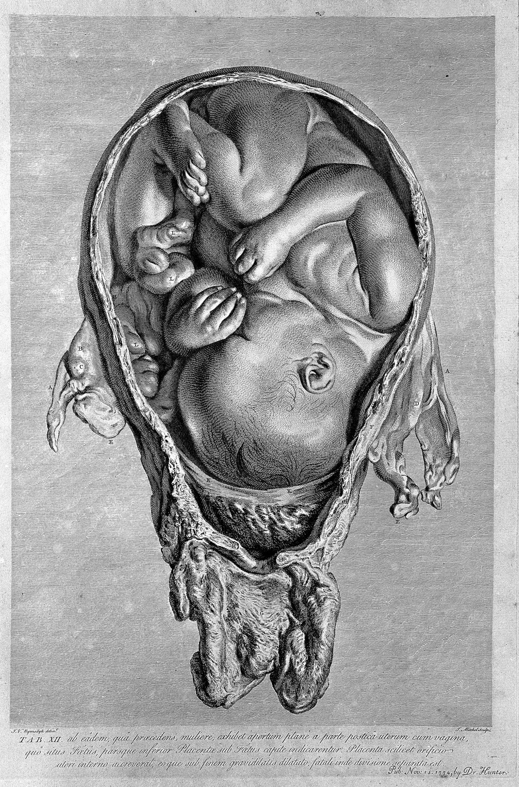
The anatomy of the human gravid uterus; foetus in utero. Wellcome L0004302
107 The anatomy of the human gravid uterus; foetus in utero. Wellcome L0004302
Unknown · CC BY 4.0 · sourceDownloadFull resolution (2692×4078) ↗
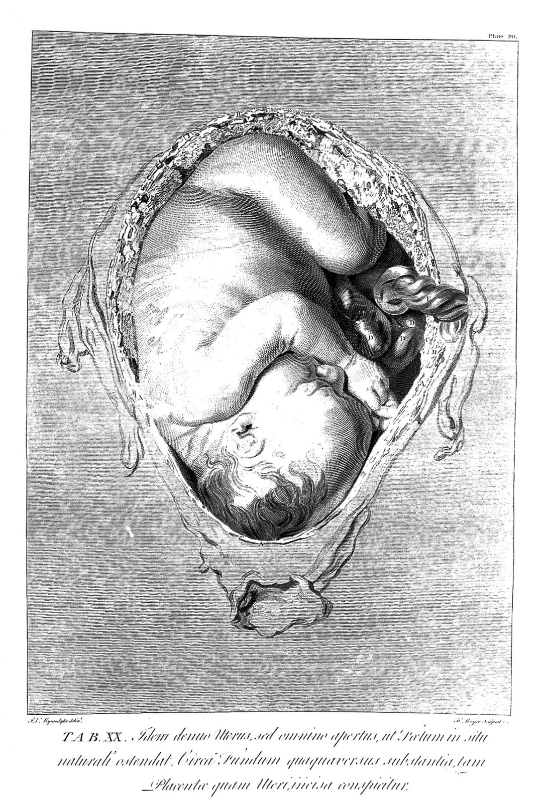
The anatomy of the human gravid uterus; foetus in utero. Wellcome L0007000
108 The anatomy of the human gravid uterus; foetus in utero. Wellcome L0007000
Unknown · CC BY 4.0 · sourceDownloadFull resolution (1152×1708) ↗
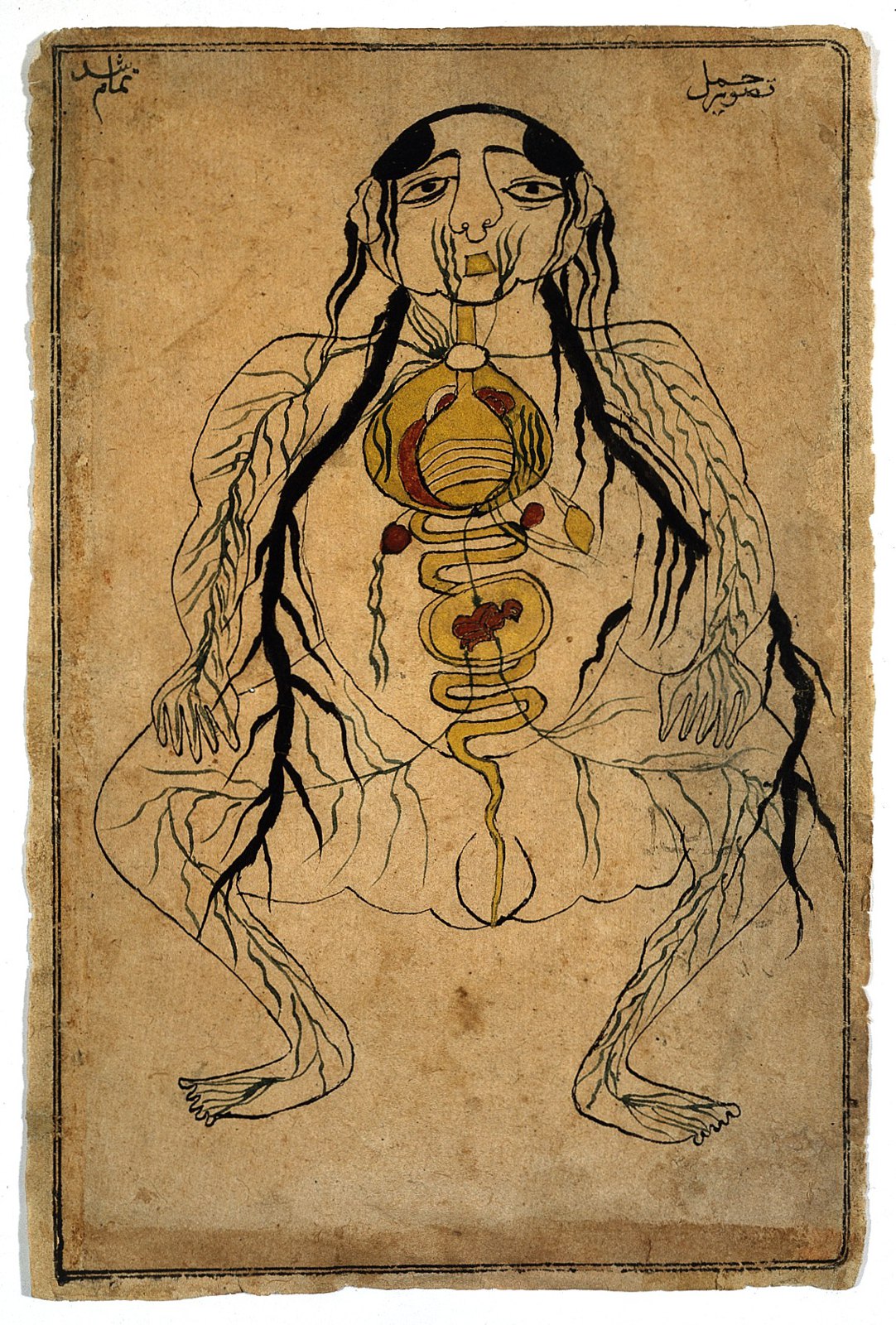
Viscera with fetus in utero, watercolour, Persian, 19th C Wellcome L0020503
109 Viscera with fetus in utero, watercolour, Persian, 19th C Wellcome L0020503
Unknown · CC BY 4.0 · sourceDownloadFull resolution (1144×1692) ↗
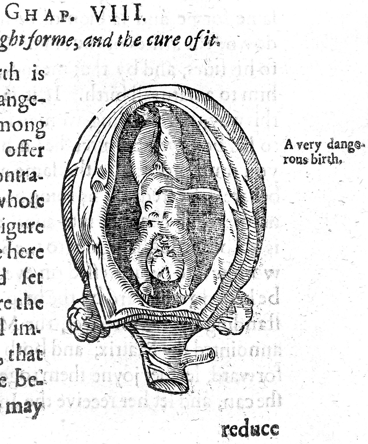
Woodcut of foetus in utero Wellcome L0014440
110 Woodcut of foetus in utero Wellcome L0014440
Unknown · CC BY 4.0 · sourceDownloadFull resolution (1275×1536) ↗
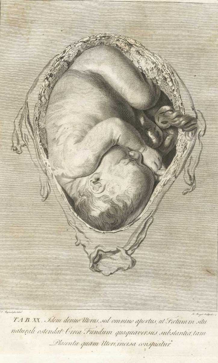
Page from "The anatomy of the human gravid uterus exhibited in figures"
111 Page from "The anatomy of the human gravid uterus exhibited in figures"
Unknown · Public domain · sourceDownloadFull resolution (724×1208) ↗
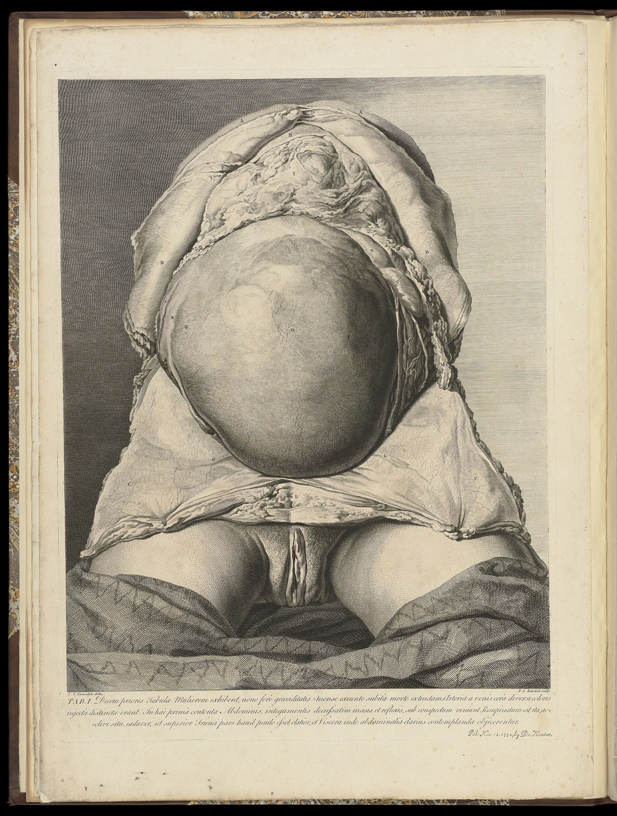
The anatomy of the human gravid uterus exhibited in figures Wellcome L0050943
112 The anatomy of the human gravid uterus exhibited in figures Wellcome L0050943
Unknown · CC BY 4.0 · sourceDownloadFull resolution (5042×6666) ↗
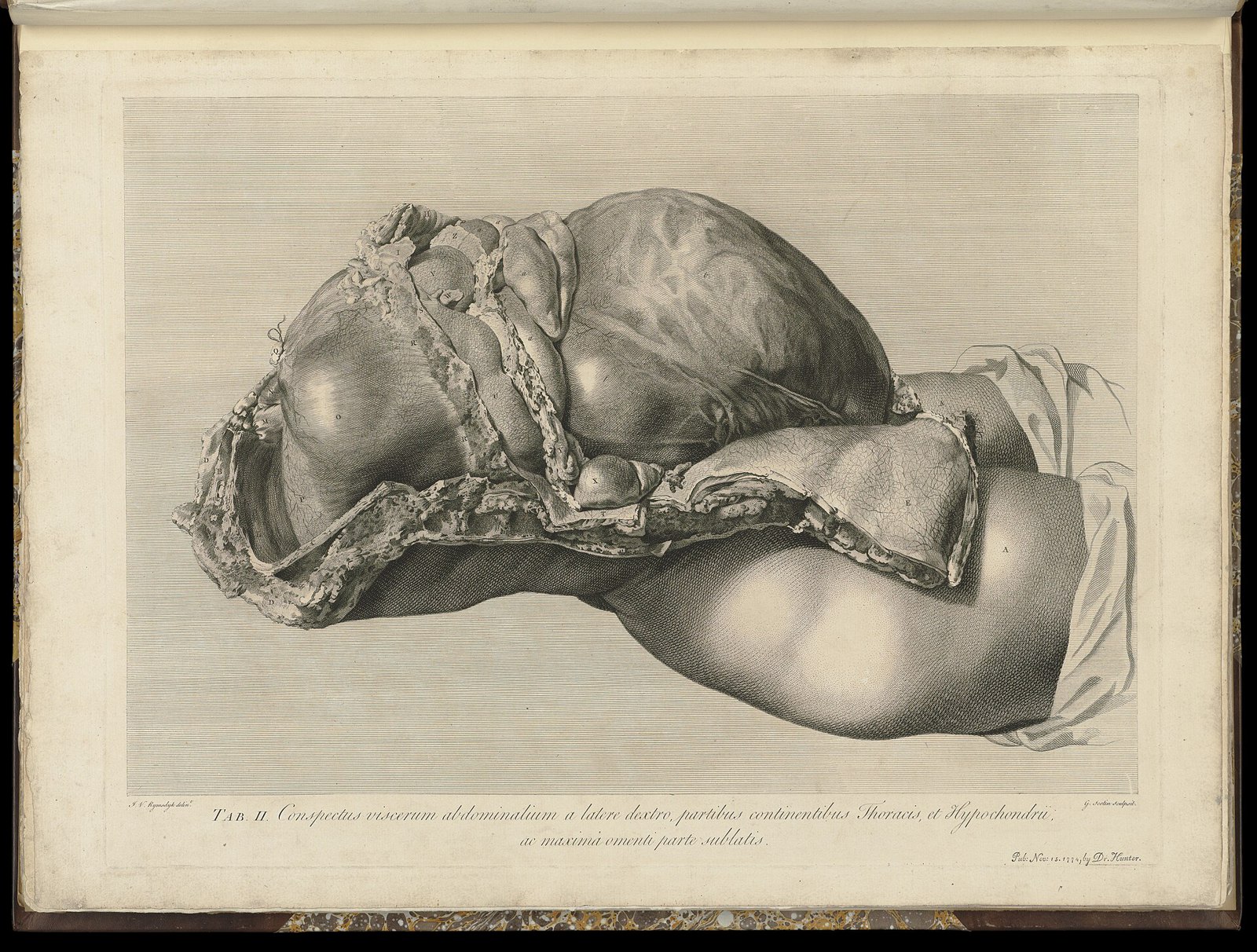
The anatomy of the human gravid uterus exhibited in figures Wellcome L0050946
113 The anatomy of the human gravid uterus exhibited in figures Wellcome L0050946
Unknown · CC BY 4.0 · sourceDownloadFull resolution (6656×5042) ↗
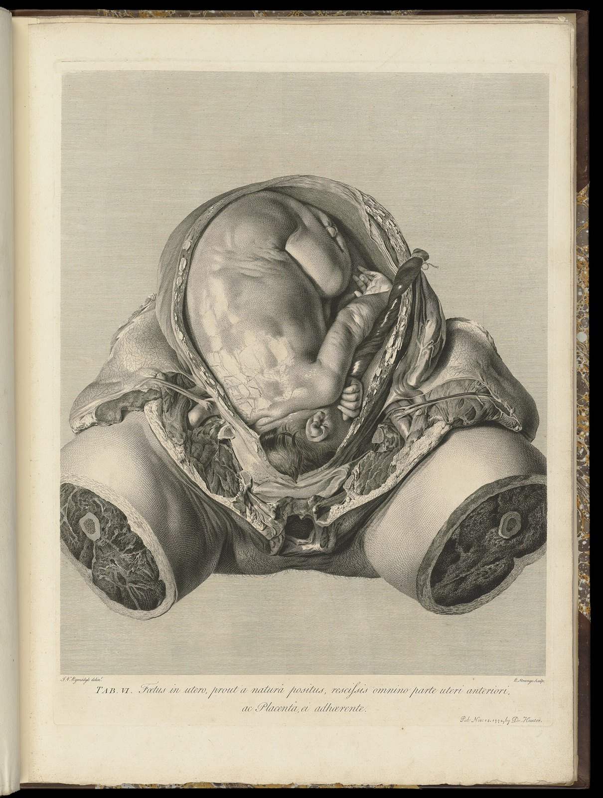
The anatomy of the human gravid uterus exhibited in figures Wellcome L0050958
114 The anatomy of the human gravid uterus exhibited in figures Wellcome L0050958
Unknown · CC BY 4.0 · sourceDownloadFull resolution (5042×6666) ↗
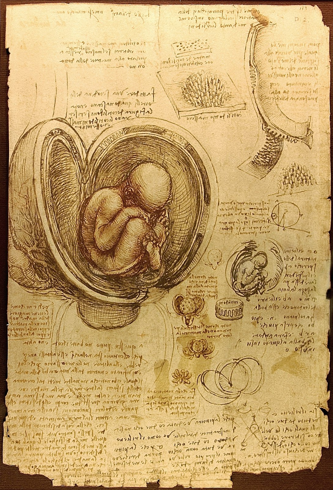
Da Vinci Studies of Embryos Luc Viatour
115 Da Vinci Studies of Embryos Luc Viatour
Leonardo da Vinci · Public domain · sourceDownloadFull resolution (1443×2121) ↗
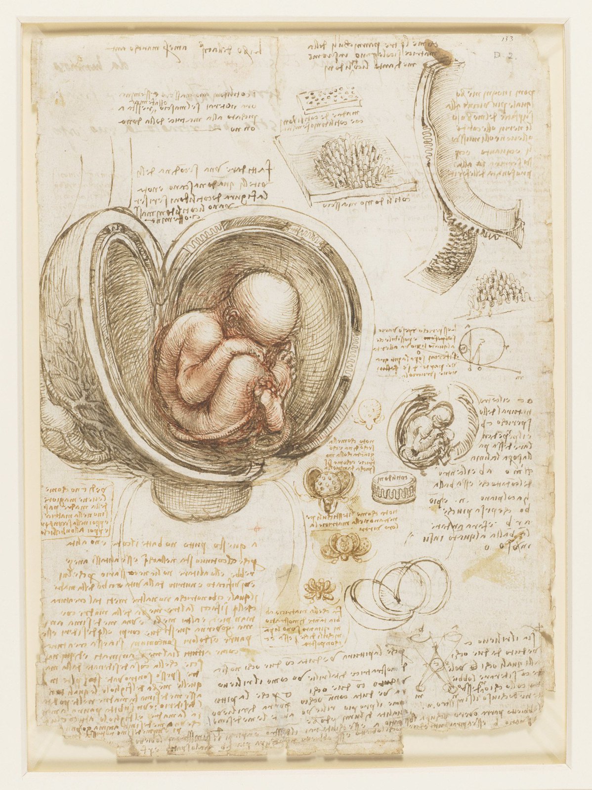
Leonardo da Vinci - RCIN 919102, Recto The fetus in the womb
116 Leonardo da Vinci - RCIN 919102, Recto The fetus in the womb
Leonardo da Vinci · Public domain · sourceDownloadFull resolution (1498×2000) ↗
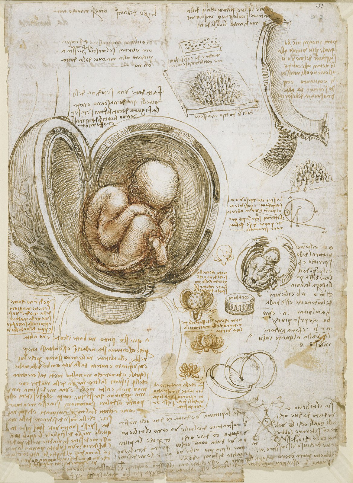
Leonardo da Vinci - Studies of the foetus in the womb
117 Leonardo da Vinci - Studies of the foetus in the womb
Leonardo da Vinci · Public domain · sourceDownloadFull resolution (2289×3136) ↗
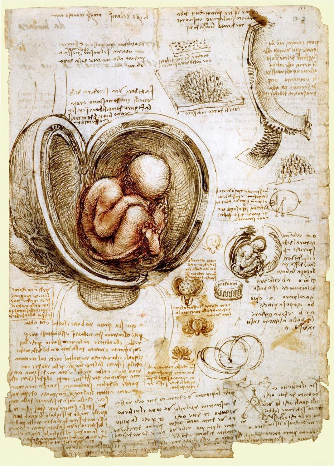
The babe in the womb; Leonardo da Vinci (1511)
118 The babe in the womb; Leonardo da Vinci (1511)
Unknown · Public domain · sourceDownloadFull resolution (1071×1492) ↗
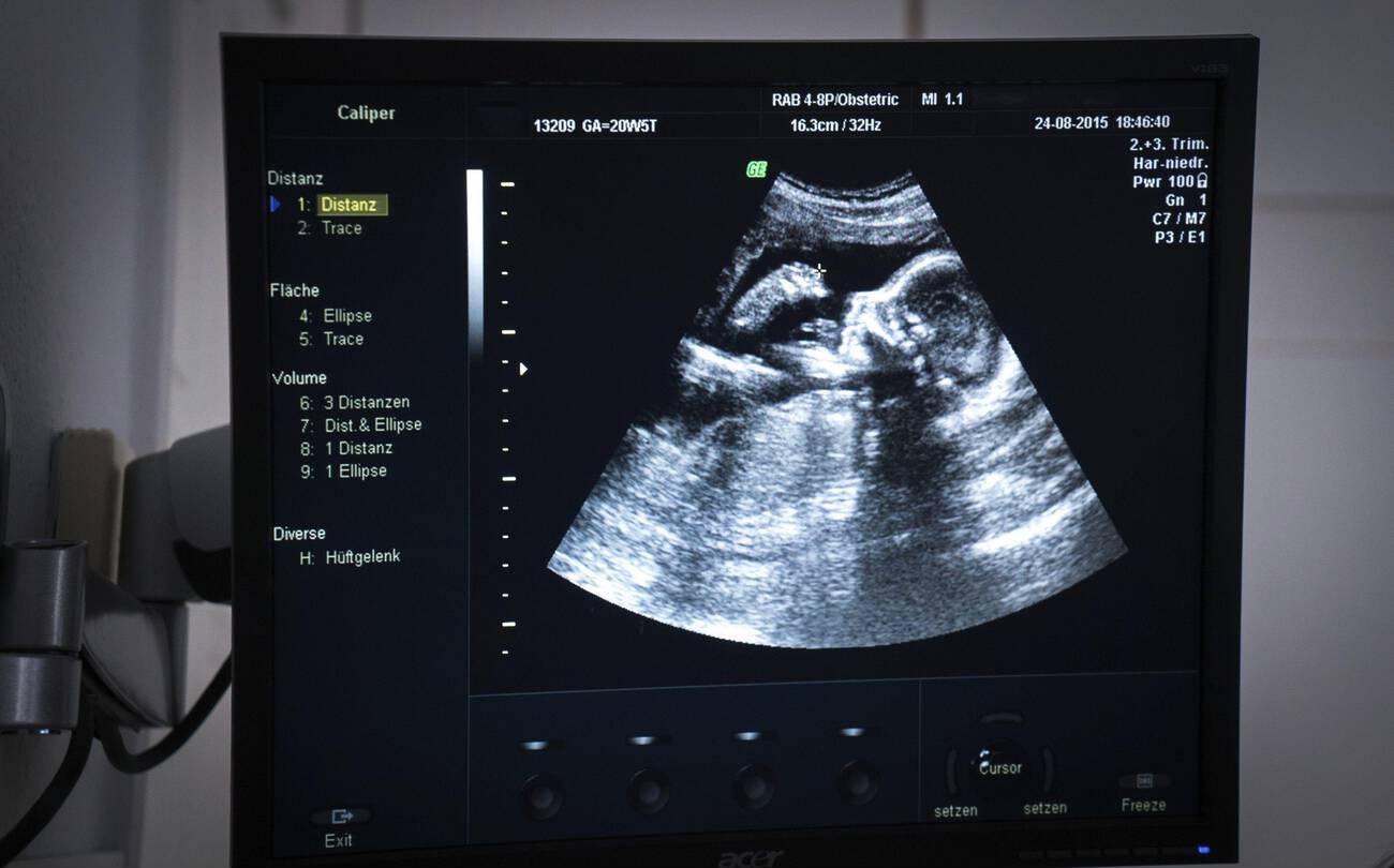
Obstetric ultrasound scan monitor showing 20-week human fetus profile in Navi Mumbai 2015
119 Obstetric ultrasound scan monitor showing 20-week human fetus profile in Navi Mumbai 2015
Goleisureintl · CC BY 4.0 · sourceDownloadFull resolution (1300×810) ↗
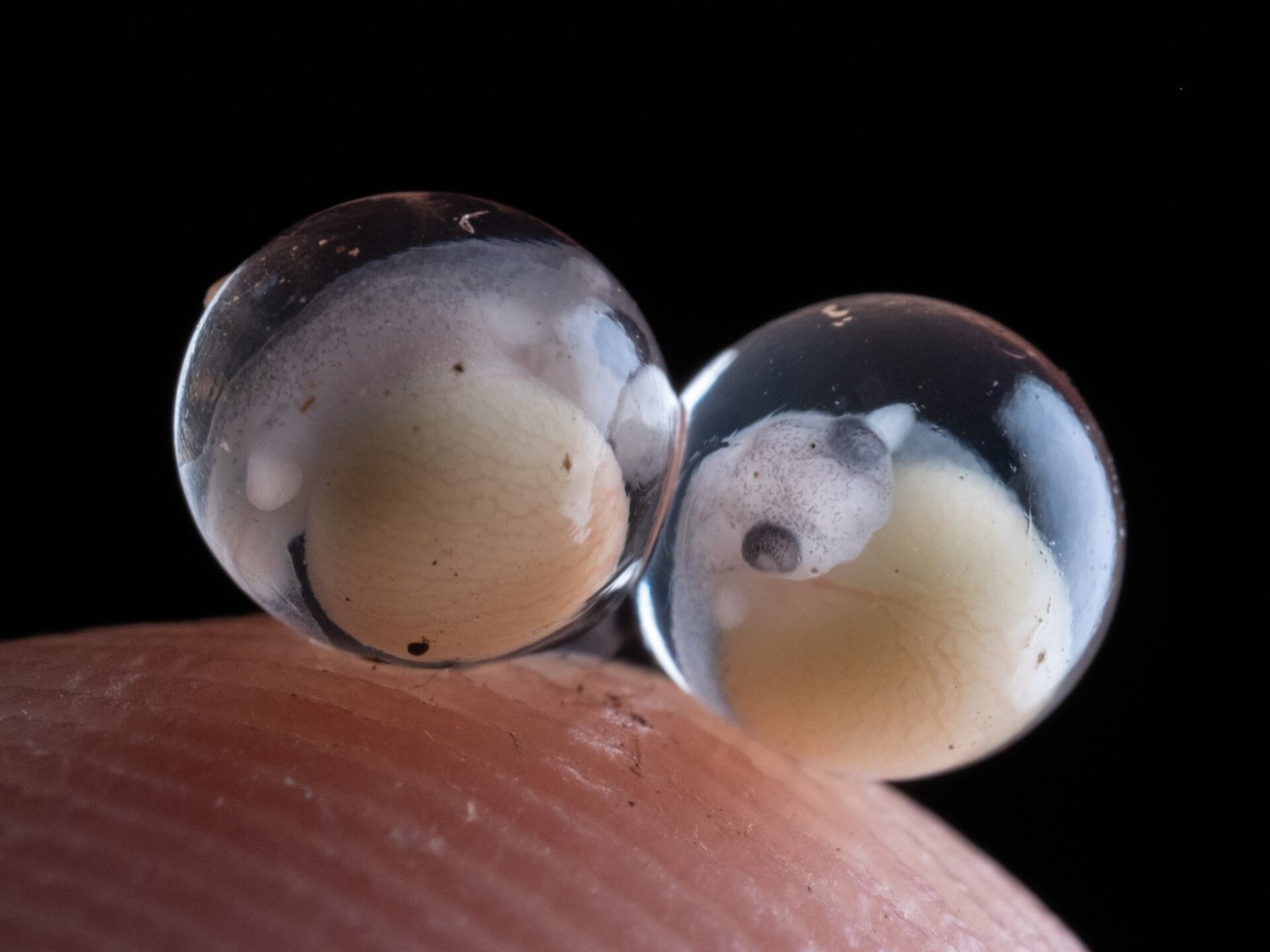
Eleutherodactylus planirostris embryos
120 Eleutherodactylus planirostris embryos
Alex Abair · CC BY 4.0 · sourceDownloadFull resolution (2048×1536) ↗
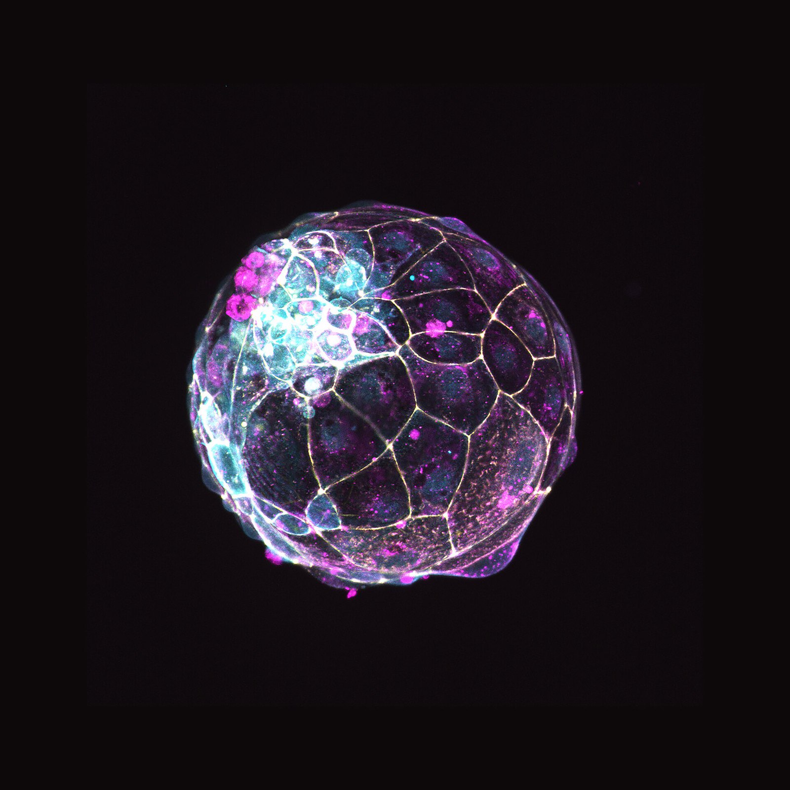
Human blastoid - 1
121 Human blastoid - 1
Nicolasrivron · CC BY-SA 4.0 · sourceDownloadFull resolution (2835×2835) ↗
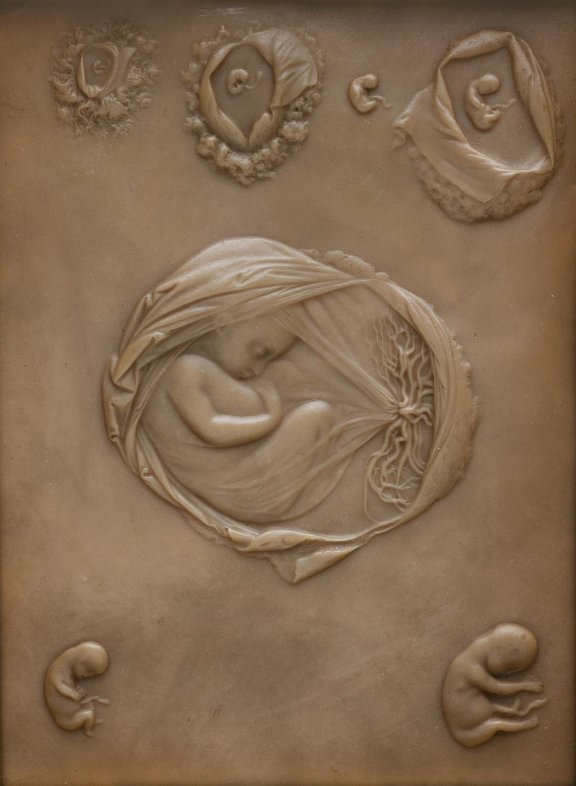
Plaque showing the development of a human embryo
122 Plaque showing the development of a human embryo
Science Museum Group Studio · CC BY-SA 4.0 · sourceDownloadFull resolution (1126×1536) ↗
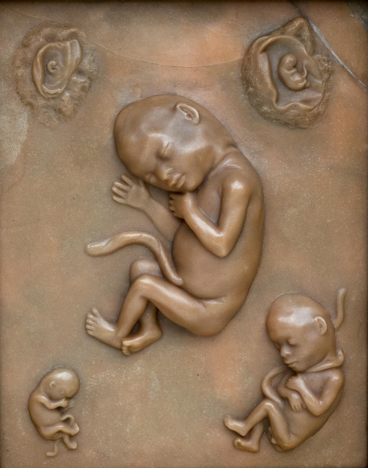
Plaque showing the development of a human embryo 2
123 Plaque showing the development of a human embryo 2
Science Museum Group Studio · CC BY-SA 4.0 · sourceDownloadFull resolution (1208×1536) ↗
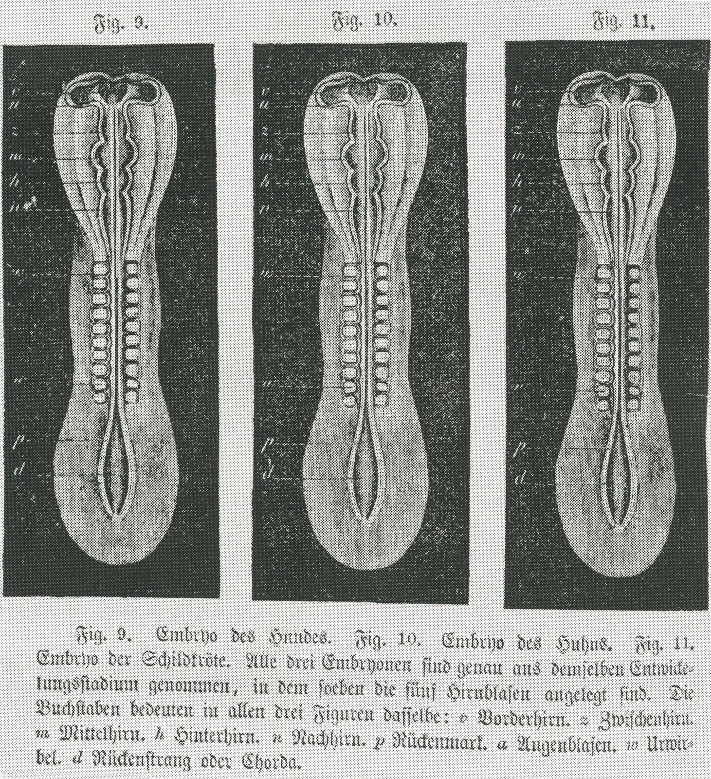
Embryo - copies
124 Embryo - copies
Creator:Ernst haeckel · Public domain · sourceDownloadFull resolution (2652×2904) ↗
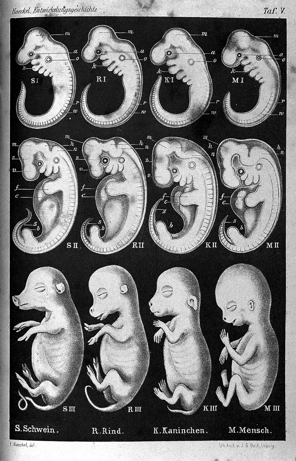
Ernst Haeckel, Anthropogenie. Wellcome L0027291
125 Ernst Haeckel, Anthropogenie. Wellcome L0027291
Unknown · CC BY 4.0 · sourceDownloadFull resolution (1102×1716) ↗
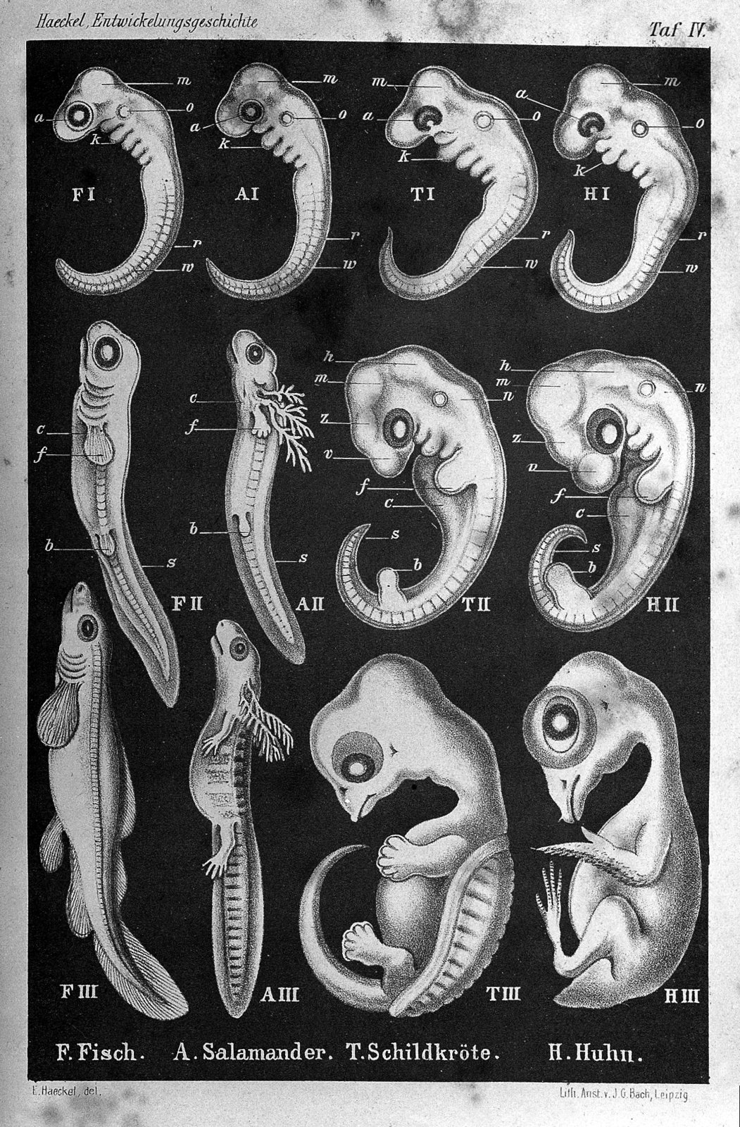
Ernst Haeckel, Anthropogenie. Wellcome L0027292
126 Ernst Haeckel, Anthropogenie. Wellcome L0027292
Unknown · CC BY 4.0 · sourceDownloadFull resolution (1360×2070) ↗
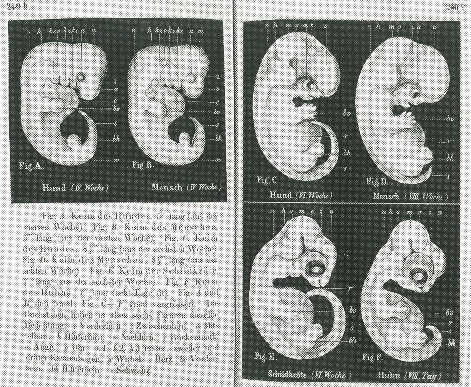
Haeckel-embryos-weeks4-6
127 Haeckel-embryos-weeks4-6
Ernst Haeckel · Public domain · sourceDownloadFull resolution (2698×2218) ↗
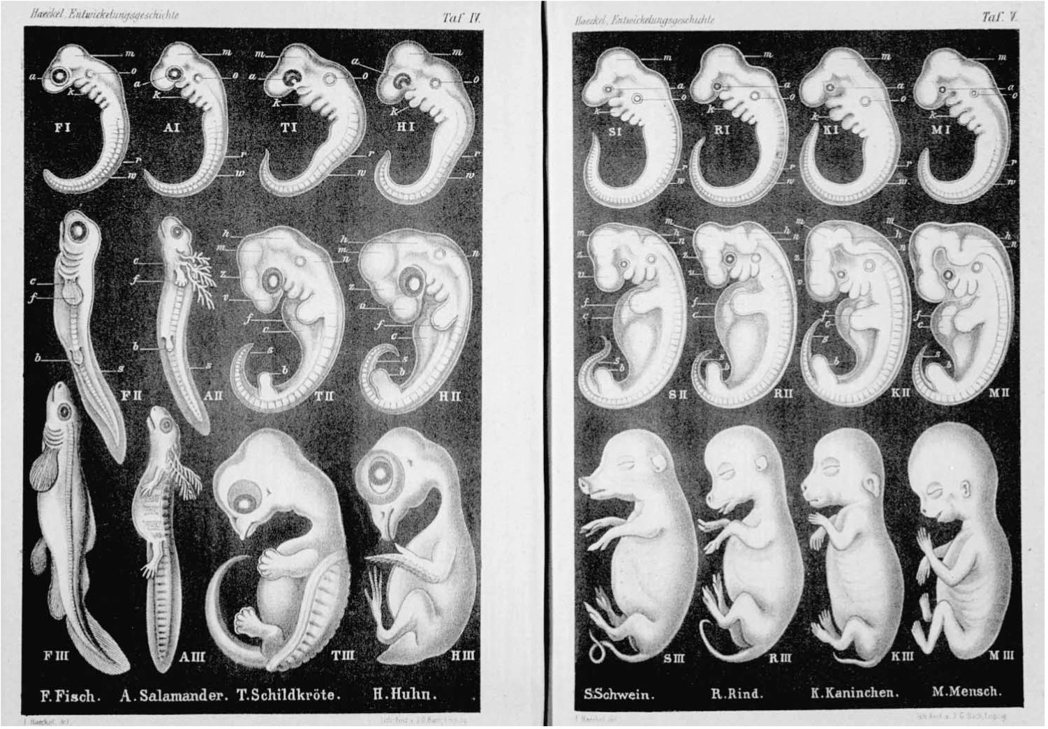
Haeckel Anthropogenie 1874
128 Haeckel Anthropogenie 1874
Lithograph by J. G. Bach of Leipzig after drawings by Haeckel, from Anthropogenie published by Engelmann · Public domain · sourceDownloadFull resolution (1501×1045) ↗
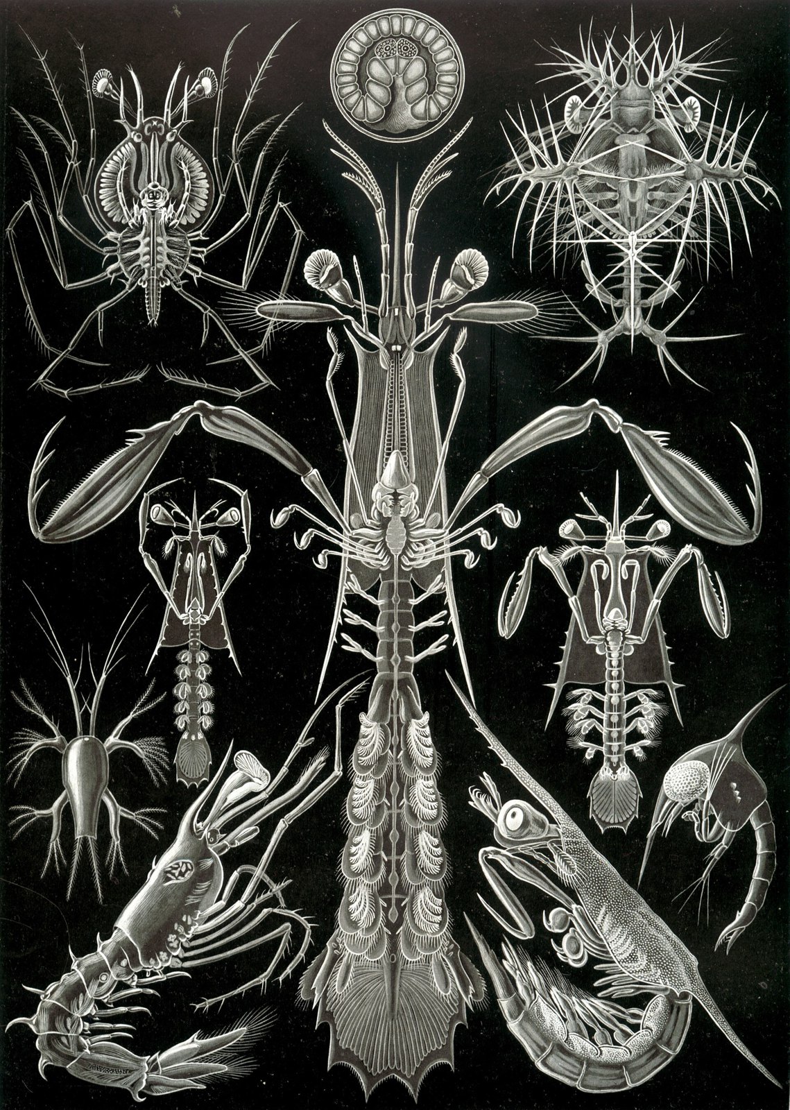
Haeckel Thoracostraca
129 Haeckel Thoracostraca
Ernst Haeckel · Public domain · sourceDownloadFull resolution (2342×3289) ↗
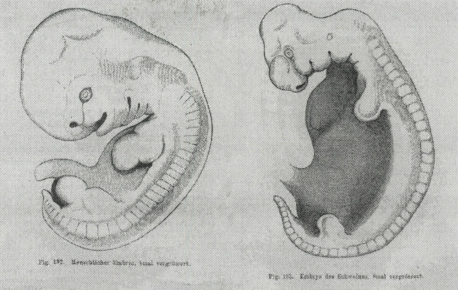
His-Embryo
130 His-Embryo
Wilhelm His · Public domain · sourceDownloadFull resolution (2704×1712) ↗
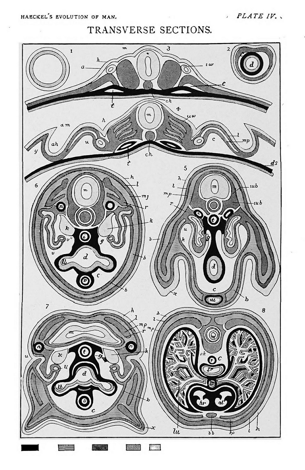
Plate IV. Diagrammatic transverse sections of different embryos
131 Plate IV. Diagrammatic transverse sections of different embryos
Haeckel, Ernst Heinrich Philipp August · Public domain · sourceDownloadFull resolution (1071×1648) ↗
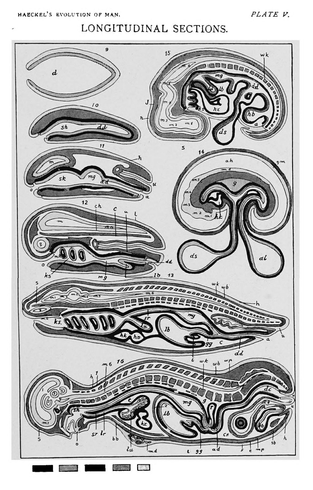
Plate V. Diagrammatic longitudinal sections of different embryos
132 Plate V. Diagrammatic longitudinal sections of different embryos
Haeckel, Ernst Heinrich Philipp August · Public domain · sourceDownloadFull resolution (1389×2174) ↗
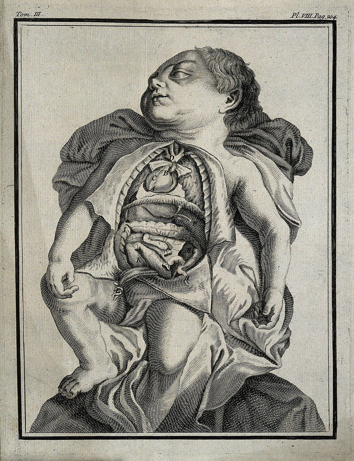
A child born with the viscera reversed. Engraving, 1749. Wellcome V0007821
133 A child born with the viscera reversed. Engraving, 1749. Wellcome V0007821
Unknown · CC BY 4.0 · sourceDownloadFull resolution (2362×3075) ↗
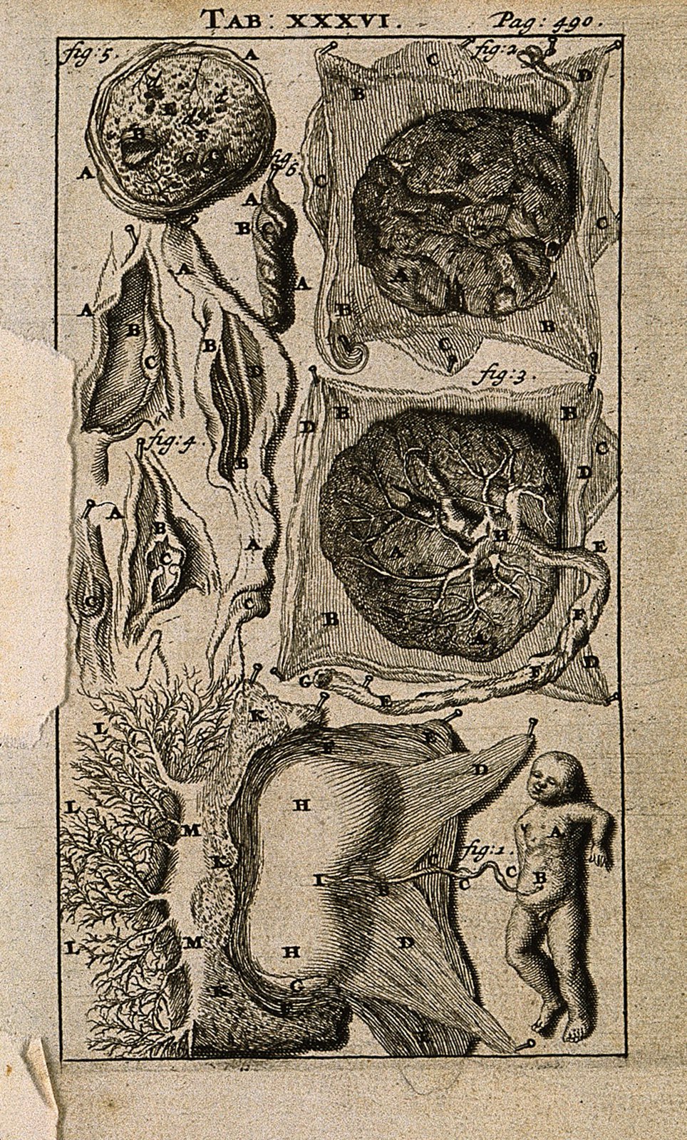
A foetus and placenta with umbilical cord. Engraving, 1686, Wellcome V0007786ER
134 A foetus and placenta with umbilical cord. Engraving, 1686, Wellcome V0007786ER
Unknown · CC BY 4.0 · sourceDownloadFull resolution (1100×1825) ↗
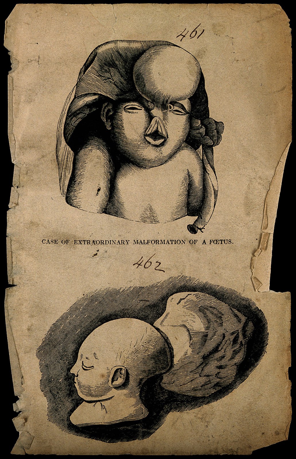
A malformed foetus. Wood engraving. Wellcome V0007401
135 A malformed foetus. Wood engraving. Wellcome V0007401
Unknown · CC BY 4.0 · sourceDownloadFull resolution (2126×3295) ↗
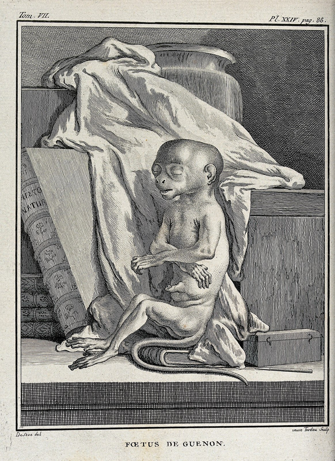
A monkey foetus. Engraving by Tardieu after De Seve. Wellcome V0021461
136 A monkey foetus. Engraving by Tardieu after De Seve. Wellcome V0021461
Unknown · CC BY 4.0 · sourceDownloadFull resolution (2300×3162) ↗
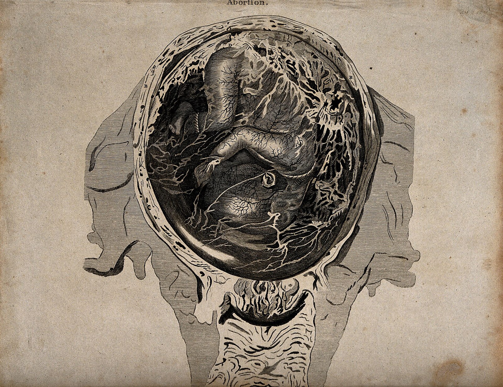
An abnormal foetus in the womb, which is about to be natural Wellcome V0014954
137 An abnormal foetus in the womb, which is about to be natural Wellcome V0014954
Unknown · CC BY 4.0 · sourceDownloadFull resolution (3136×2412) ↗
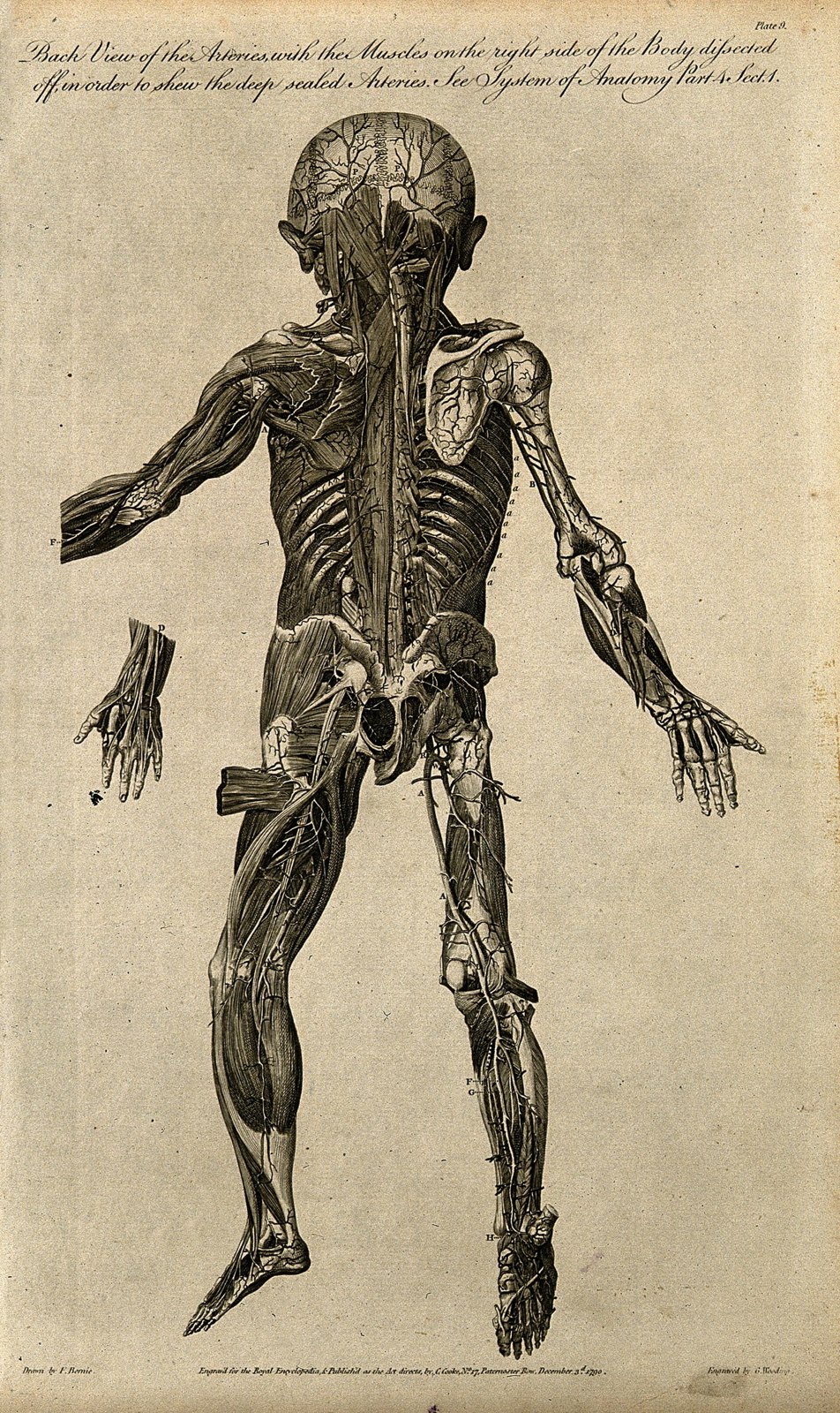
Dissected foetus seen from behind, with arteries and muscles Wellcome V0007991
138 Dissected foetus seen from behind, with arteries and muscles Wellcome V0007991
Unknown · CC BY 4.0 · sourceDownloadFull resolution (2125×3573) ↗
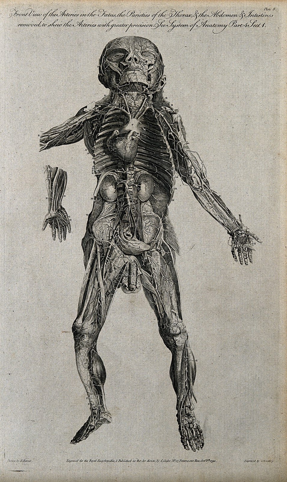
Dissected foetus seen from the front, with arteries indicate Wellcome V0007990
139 Dissected foetus seen from the front, with arteries indicate Wellcome V0007990
Unknown · CC BY 4.0 · sourceDownloadFull resolution (2050×3434) ↗
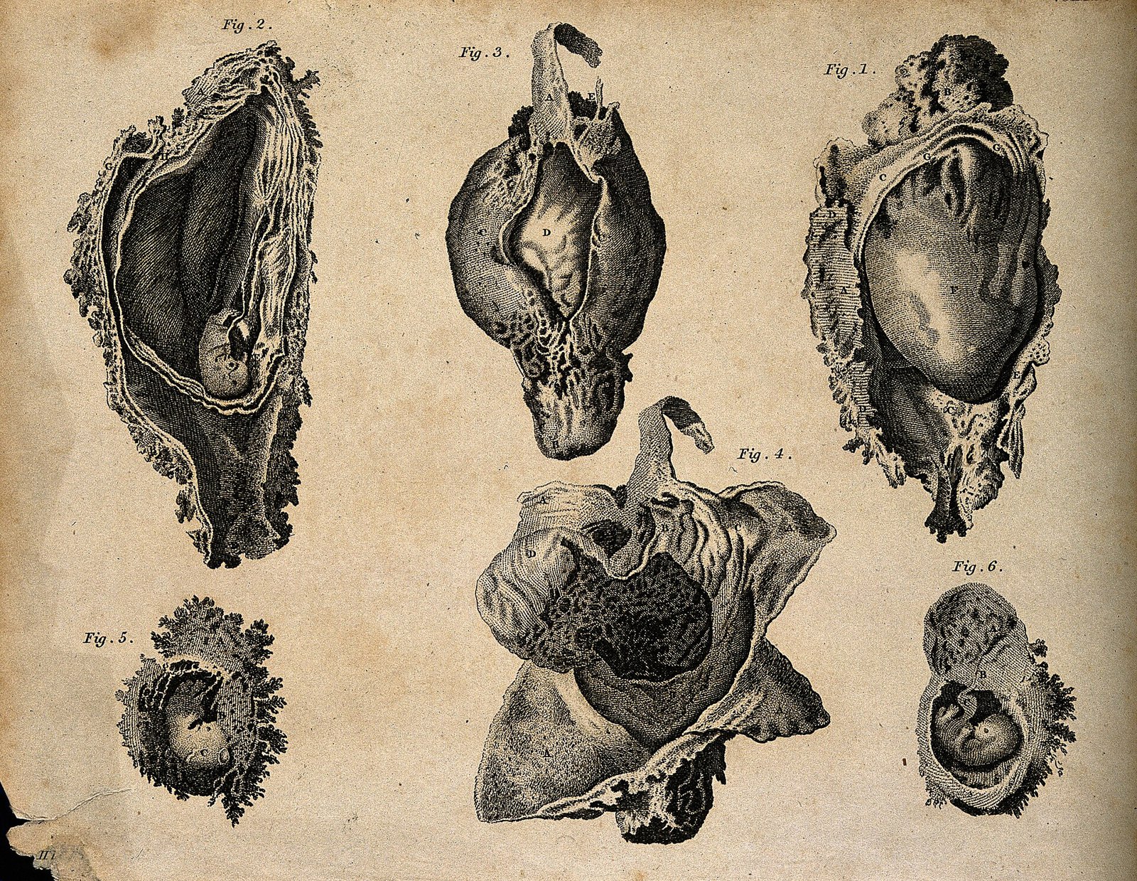
Foetus in utero; six figures. Stipple engraving by Campbell( Wellcome V0008049
140 Foetus in utero; six figures. Stipple engraving by Campbell( Wellcome V0008049
Unknown · CC BY 4.0 · sourceDownloadFull resolution (3124×2420) ↗
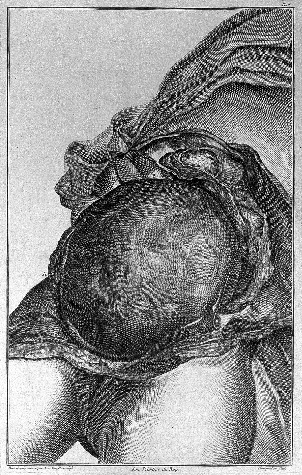
Jenty; anatomy of the womb and foetus Wellcome L0030022
141 Jenty; anatomy of the womb and foetus Wellcome L0030022
Unknown · CC BY 4.0 · sourceDownloadFull resolution (1176×1851) ↗
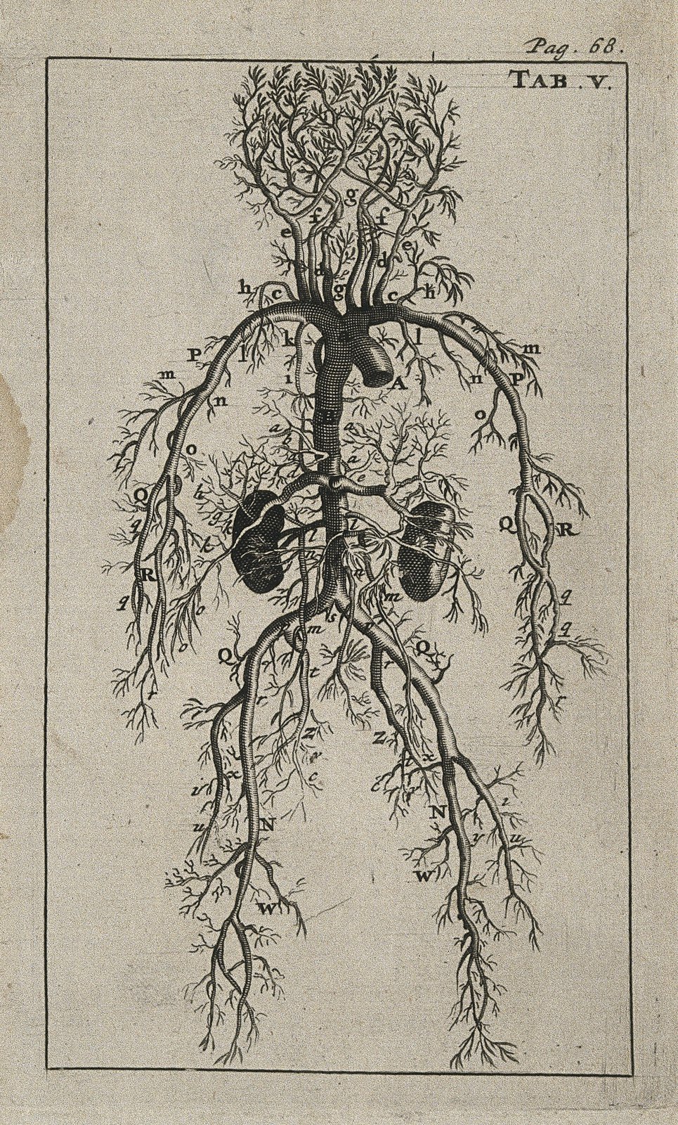
The arterial system of human foetus. Engraving, 1686, after Wellcome V0007779EC
142 The arterial system of human foetus. Engraving, 1686, after Wellcome V0007779EC
Unknown · CC BY 4.0 · sourceDownloadFull resolution (2515×4172) ↗
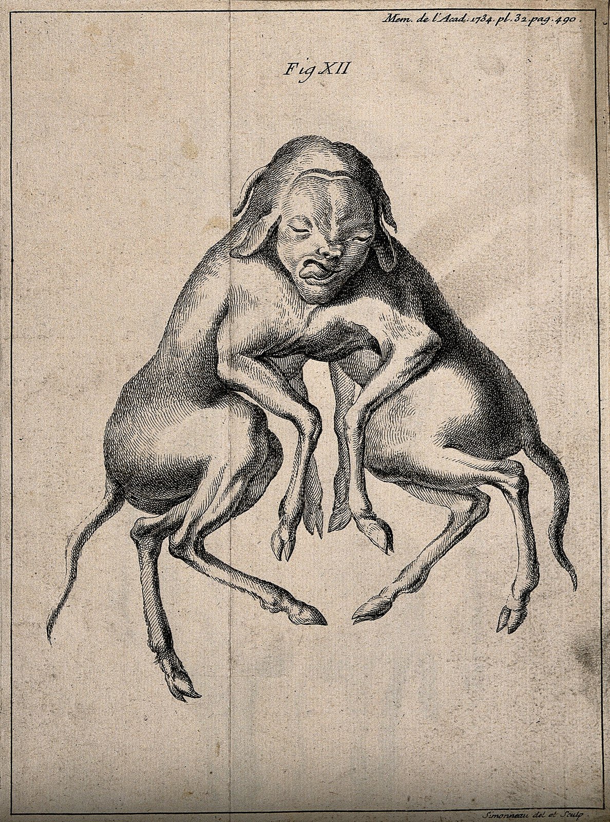
The foetus of dogs born as conjoint twins, anterior view. En Wellcome V0007390
143 The foetus of dogs born as conjoint twins, anterior view. En Wellcome V0007390
Unknown · CC BY 4.0 · sourceDownloadFull resolution (2284×3076) ↗
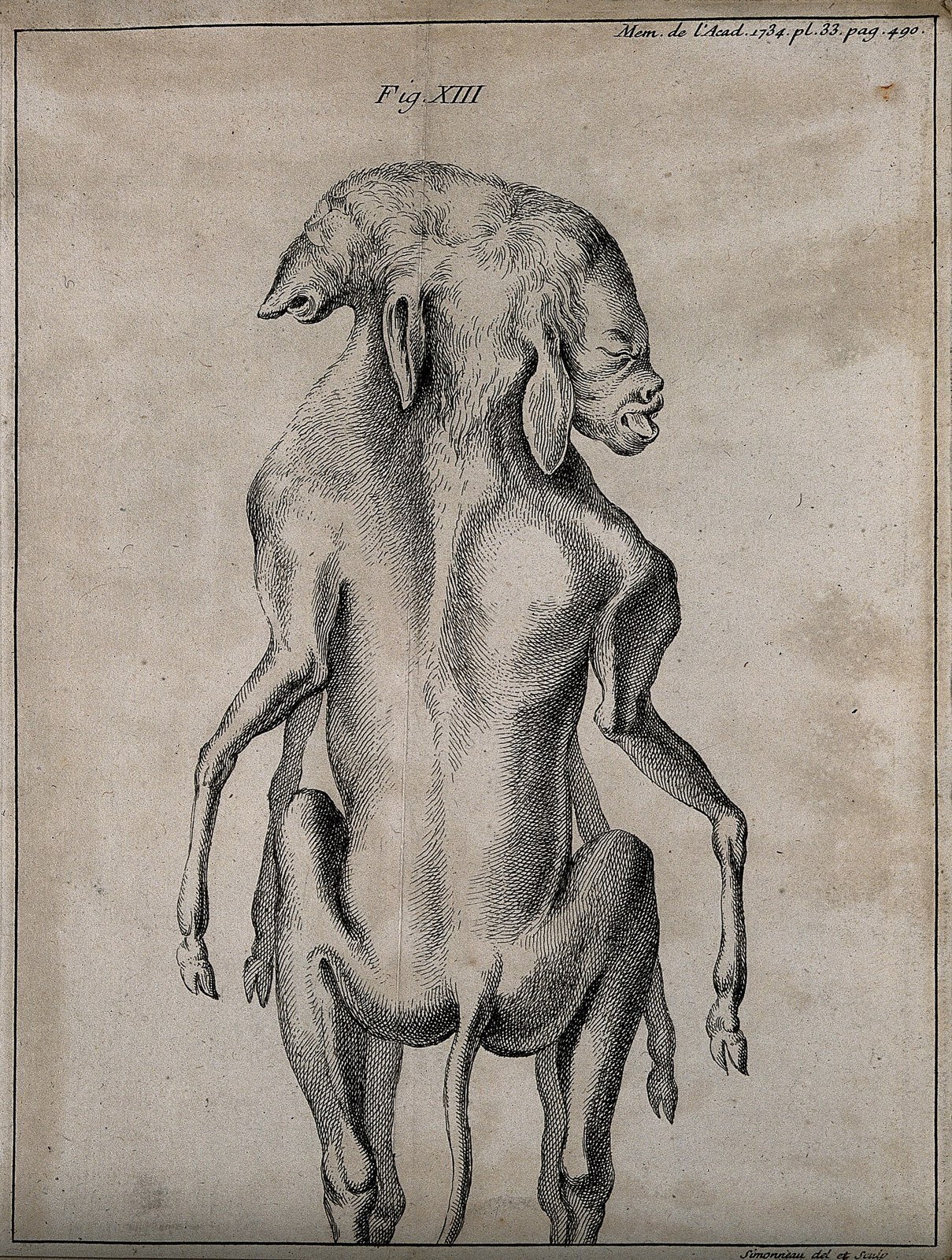
The foetus of dogs born as conjoint twins. Engraving by P. S Wellcome V0007391
144 The foetus of dogs born as conjoint twins. Engraving by P. S Wellcome V0007391
Unknown · CC BY 4.0 · sourceDownloadFull resolution (2394×3167) ↗
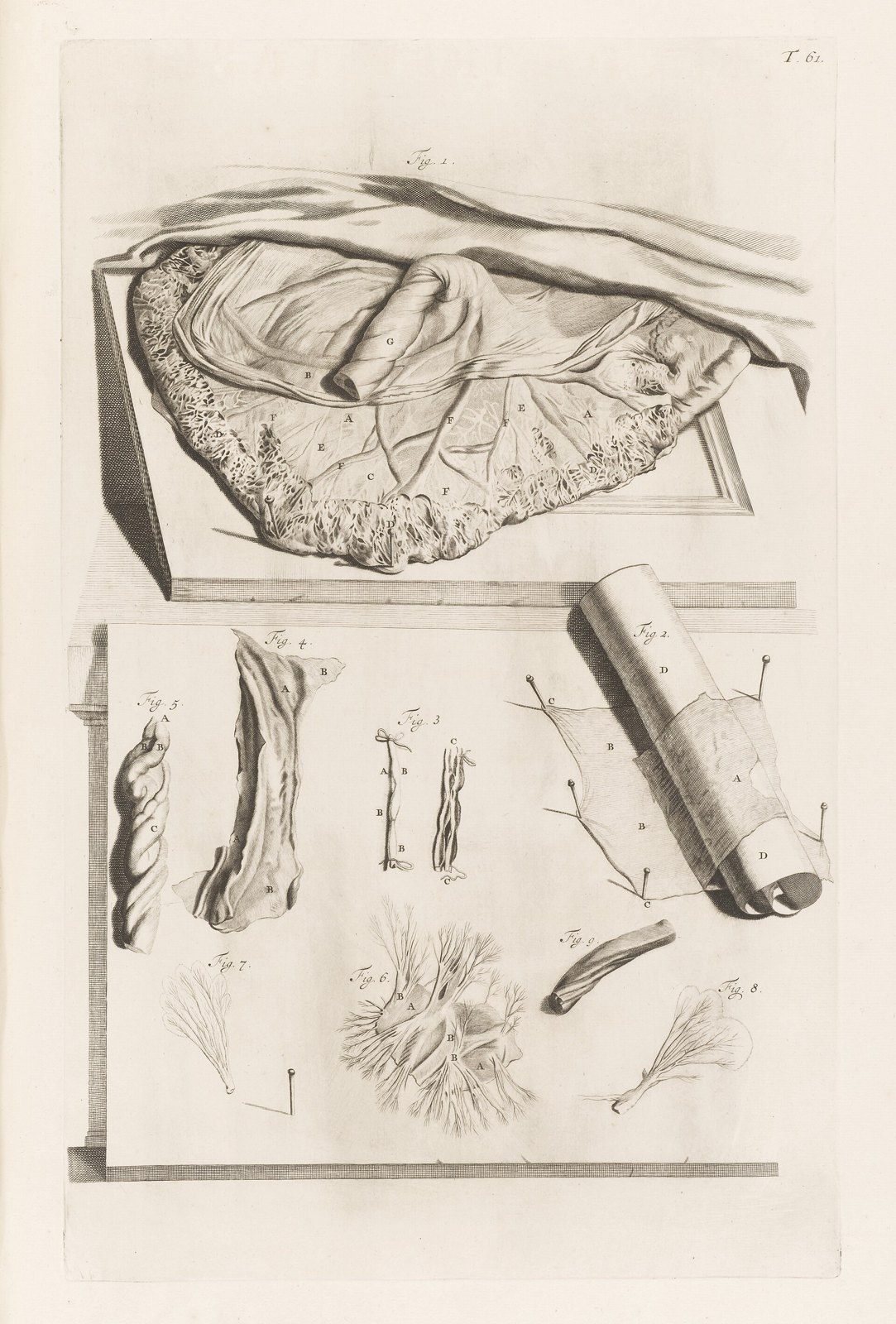
Illustration of a placenta Wellcome L0051128
145 Illustration of a placenta Wellcome L0051128
Unknown · CC BY 4.0 · sourceDownloadFull resolution (4477×6613) ↗
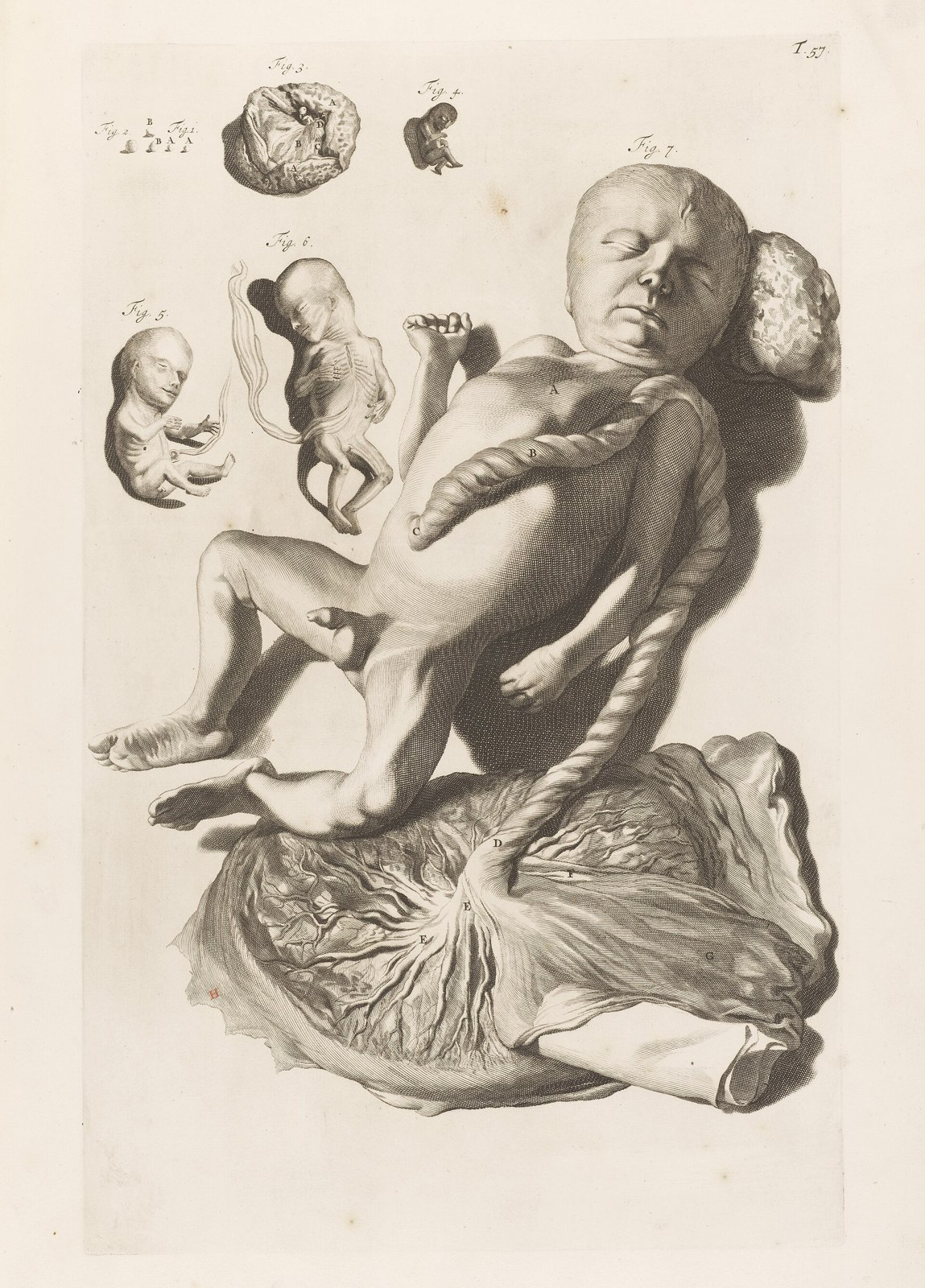
Illustration of male foetus with placenta Wellcome L0051127
146 Illustration of male foetus with placenta Wellcome L0051127
Unknown · CC BY 4.0 · sourceDownloadFull resolution (4772×6643) ↗
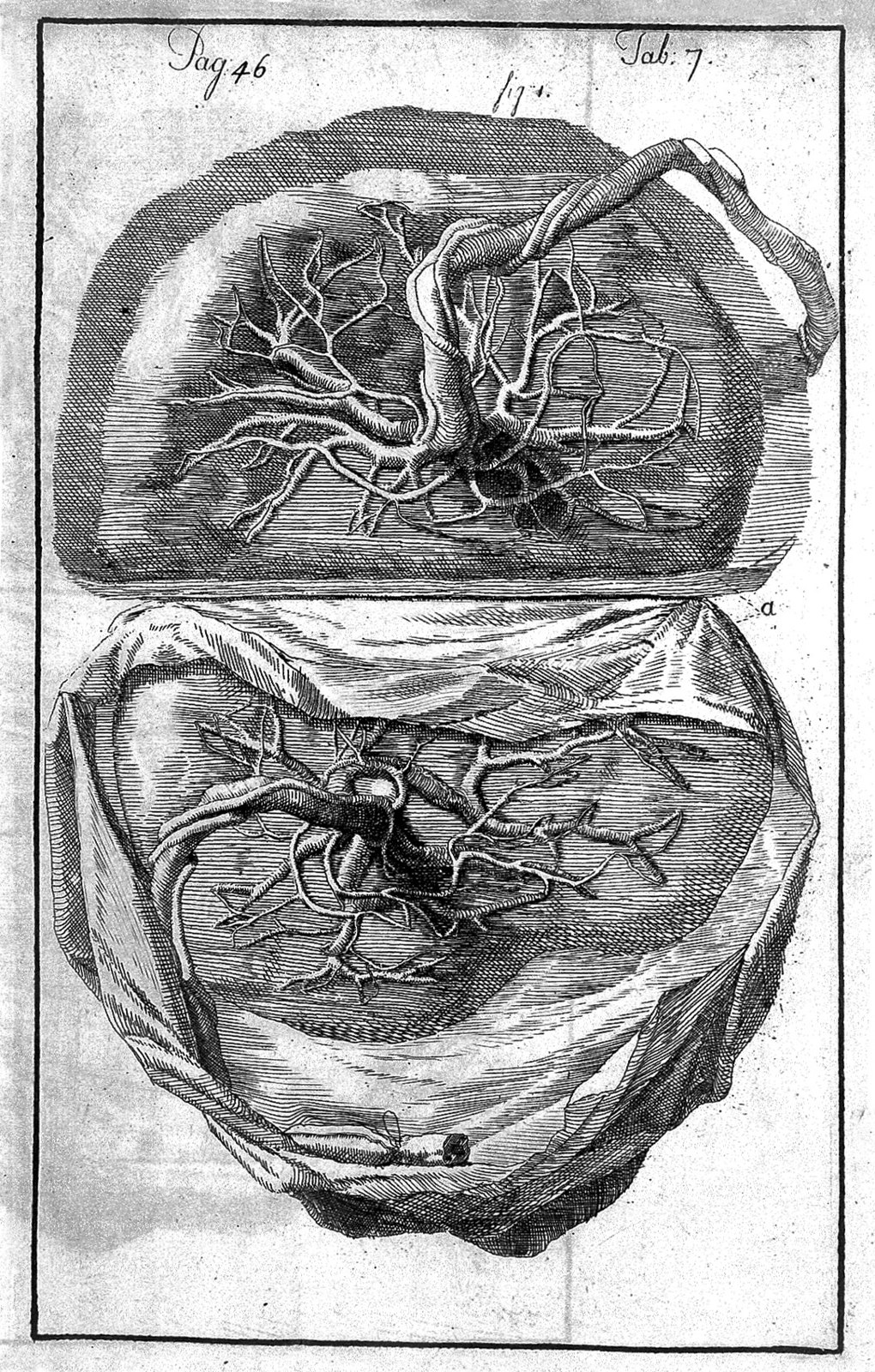
Placenta belonging to twins. Wellcome L0009816
147 Placenta belonging to twins. Wellcome L0009816
Unknown · CC BY 4.0 · sourceDownloadFull resolution (1084×1701) ↗
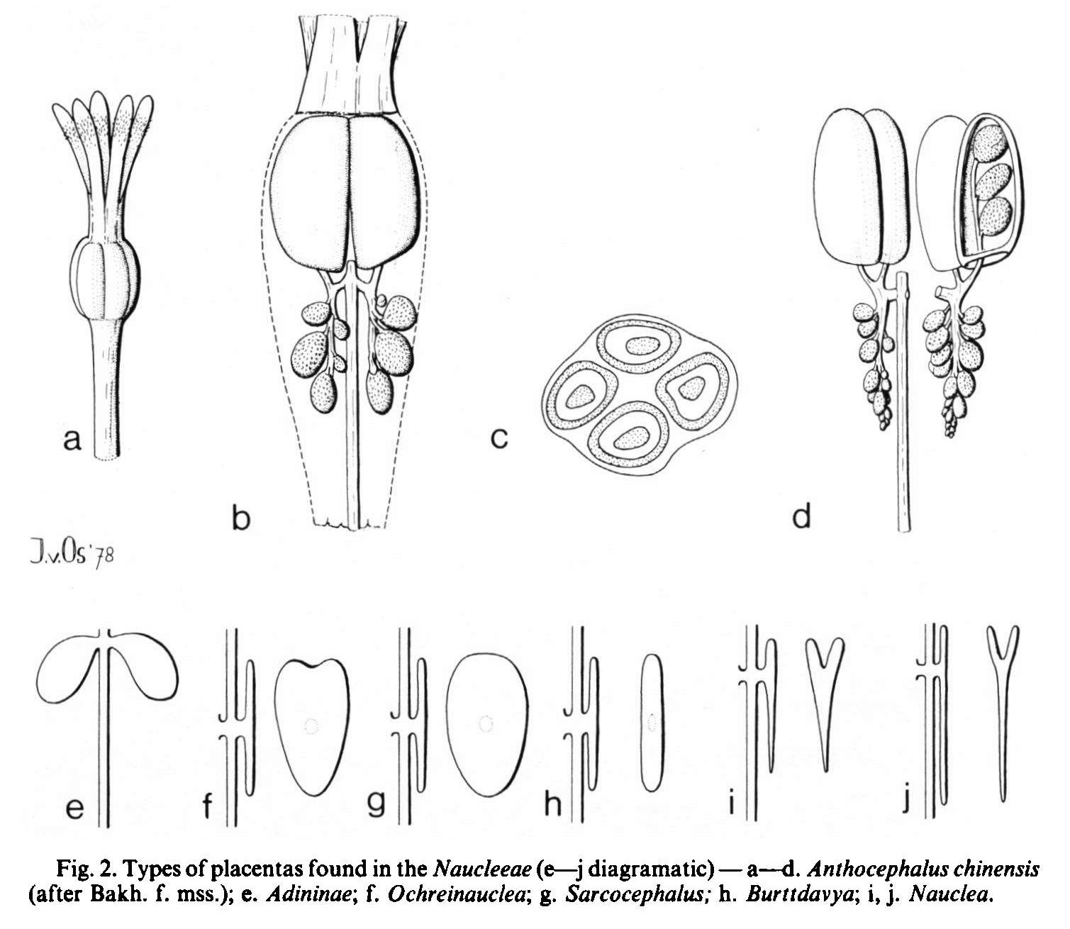
Placenta types in Naucleeae from Blumea 24(2) 315 (1978)
148 Placenta types in Naucleeae from Blumea 24(2) 315 (1978)
Illlustration: J. van Os · CC BY 4.0 · sourceDownloadFull resolution (1509×1310) ↗
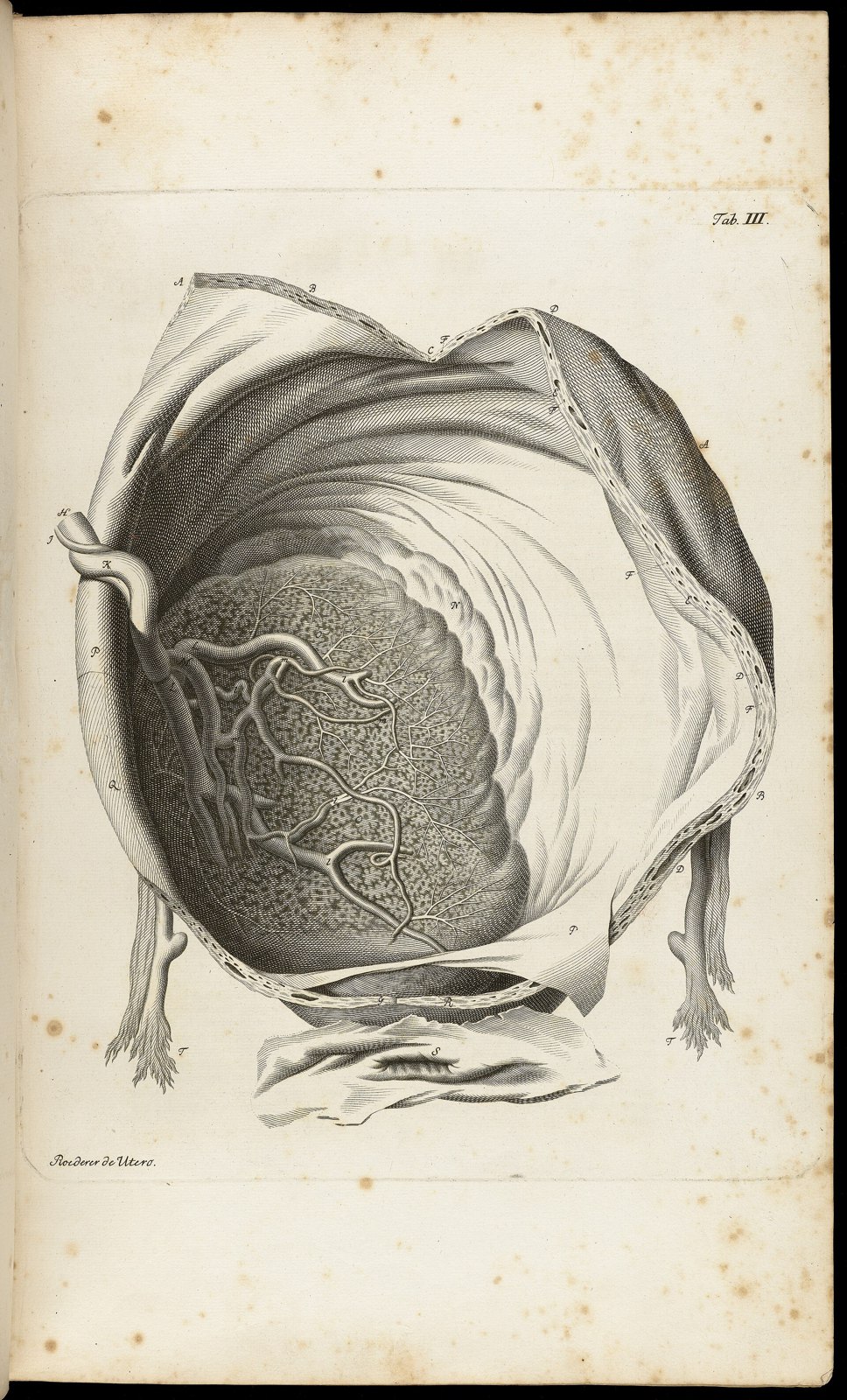
Tab III; Placenta within amneotic sac Wellcome L0064774
149 Tab III; Placenta within amneotic sac Wellcome L0064774
Unknown · CC BY 4.0 · sourceDownloadFull resolution (3854×6370) ↗
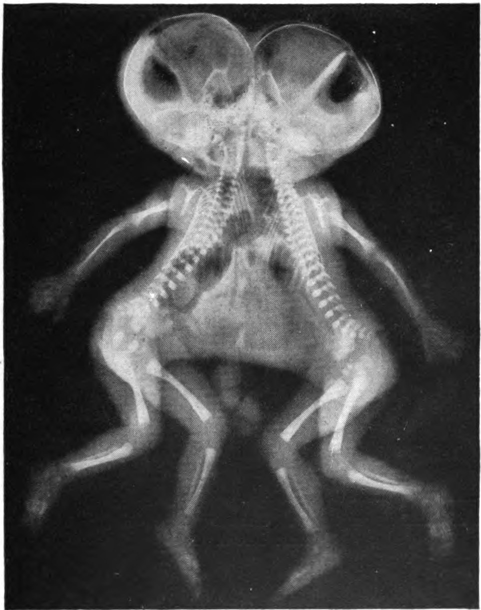
Conjoined X-ray
150 Conjoined X-ray
Mae M. Bookmiller · Public domain · sourceDownloadFull resolution (963×1220) ↗
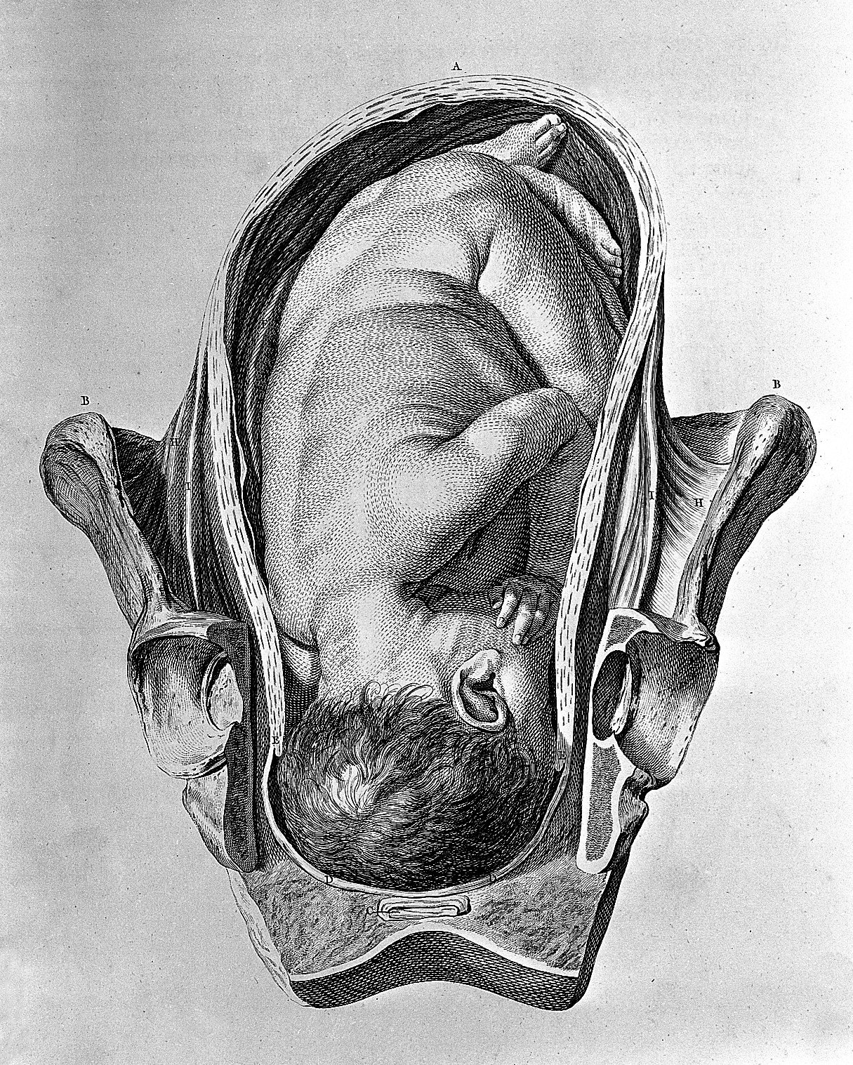
Foetus in utero. Wellcome L0004300
151 Foetus in utero. Wellcome L0004300
Unknown · CC BY 4.0 · sourceDownloadFull resolution (1215×1517) ↗
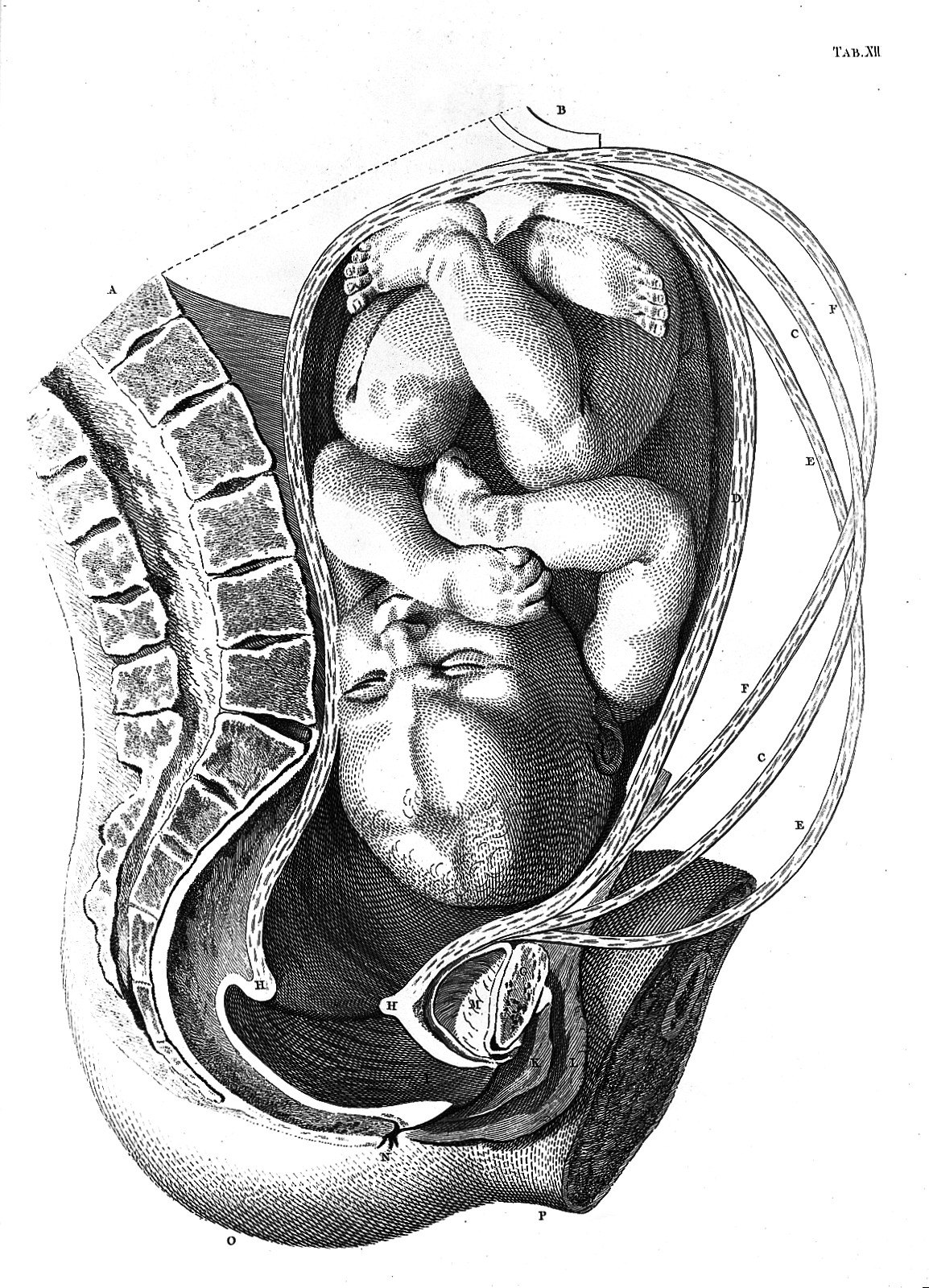
Foetus in utero. Wellcome L0013508
152 Foetus in utero. Wellcome L0013508
Unknown · CC BY 4.0 · sourceDownloadFull resolution (1154×1600) ↗
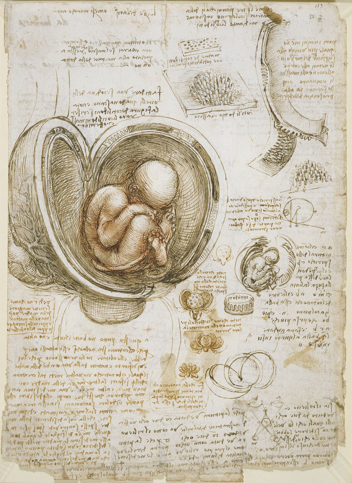
Leonardo da Vinci - Studies of the foetus in the womb
153 Leonardo da Vinci - Studies of the foetus in the womb
Leonardo da Vinci · Public domain · sourceDownloadFull resolution (2289×3136) ↗